What should I do now?
Call 911
Call 911 for chest pressure, severe trouble breathing, fainting during a racing heartbeat, or stroke signs such as face drooping, arm weakness, or trouble speaking. Do not drive yourself.
Contact your care team
Contact your care team for new or longer episodes, worsening fatigue, reduced activity, medication side effects, or bleeding concerns.
Keep your care plan
Keep your treatment and follow-up plan. Bring questions, readings, and your medication list.
04 / TREATMENT
4. How is AFib treated?
AFib treatment addresses several different goals: protecting you from stroke, preventing an excessively fast heartbeat, and reducing AFib episodes. Your treatment plan is tailored to your symptoms, medical conditions, and personal goals.
The three pillars of AFib treatment
Each pillar has a different job. A medication that slows your heart rate does not replace stroke prevention, and successful rhythm treatment does not automatically mean you can stop a blood thinner.
The foundation: treating contributing risk factors
Alongside these three pillars, we address conditions and habits that contribute to AFib, such as:
- High blood pressure.
- Excess weight.
- Sleep apnea.
- Alcohol use.
- Smoking.
- Physical inactivity.
- Diabetes and other heart conditions.
Treating these risk factors does not cure AFib, but it can reduce symptoms and decrease the frequency and duration of episodes. It can also help other AFib treatments work better.
Your plan may combine several treatments, with adjustments over time as your needs change.
Pillar 1: Preventing blood clots and stroke
Not everyone with AFib has the same stroke risk. We use a guideline-recommended stroke risk score, commonly called CHA₂DS₂-VASc, to estimate your likelihood of having a stroke. It considers your age and conditions such as high blood pressure, diabetes, heart failure, vascular disease, and a previous stroke.
Your score helps guide treatment, along with your other medical conditions and bleeding risk. We reassess it over time because your risk can change.
When do I need a blood thinner?
If your stroke risk is high enough, we usually recommend an anticoagulant, often called a blood thinner.
These medications do not actually make blood thinner. They reduce the blood’s ability to form clots, making it less likely that a clot will form in the left atrial appendage and travel to the brain. This lowers the risk of an ischemic stroke—a stroke caused by a blocked artery.
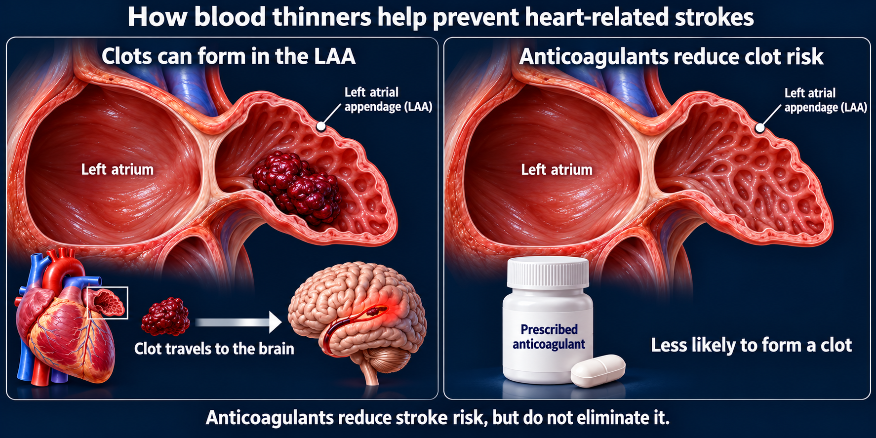
Blood thinners help prevent strokes, but they do not stop AFib or control your heart rate. They also increase bleeding risk, which we consider when choosing your treatment.
What if I cannot tolerate a blood thinner?
Some patients need stroke protection but cannot safely take a blood thinner long term—for example, because of serious bleeding.
For selected patients, we may recommend left atrial appendage closure. During this procedure, a device such as Watchman or Amulet is placed inside the appendage to seal off the pouch where AFib-related clots commonly form.
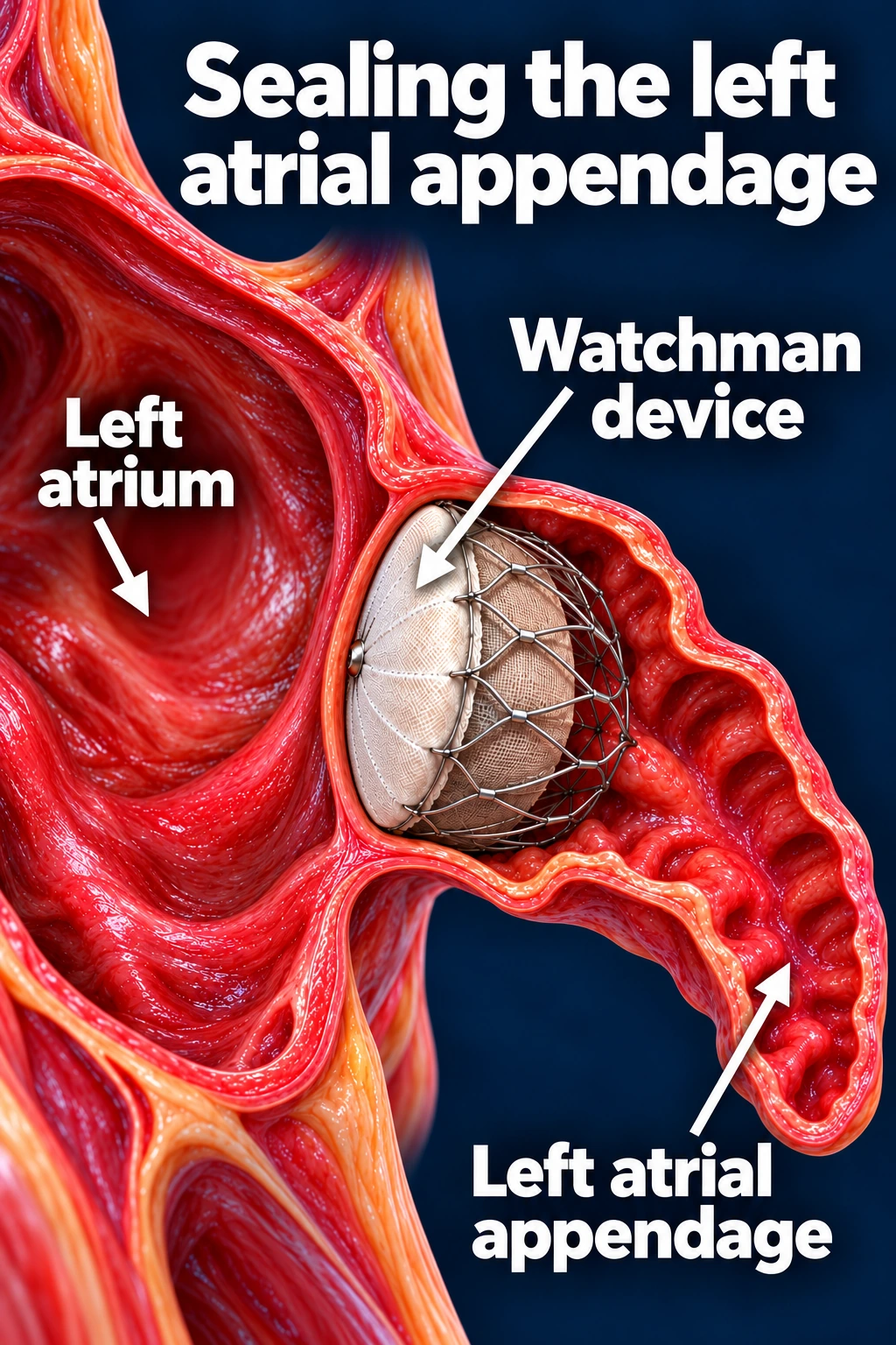
This can provide an alternative to long-term blood thinners for appropriate patients. It does not treat AFib itself, and blood-thinning or antiplatelet medication is usually needed for a period after implantation while the device heals.
The goal is to choose a stroke-prevention plan that balances protection from clots with your individual bleeding risk.
Pillar 2: Controlling the heart rate
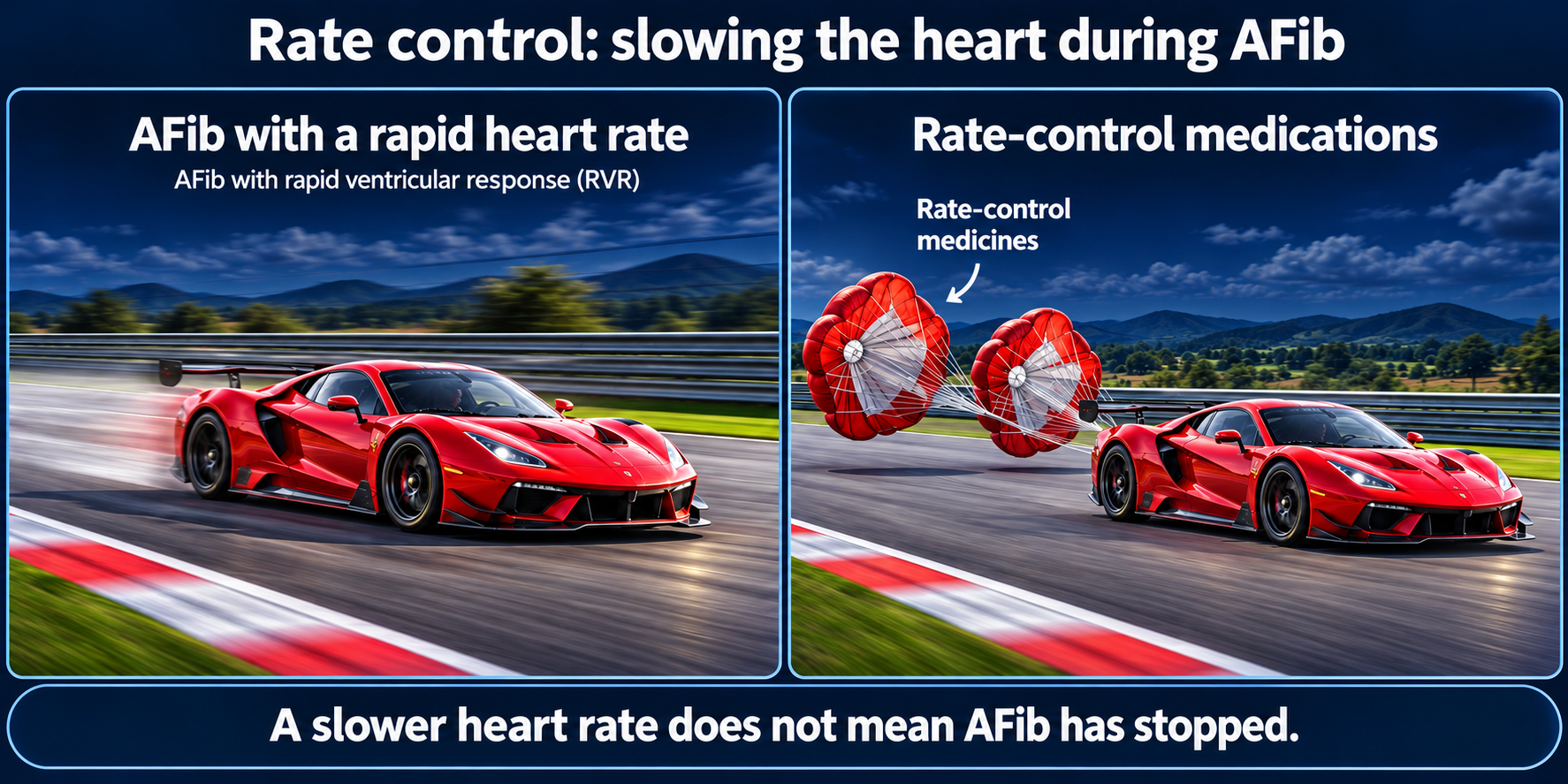
During AFib, the disorganized signals in the upper chambers can cause the lower chambers to beat too quickly. Rate-control medications slow this response, helping prevent an excessively fast pulse.
These medications do not stop the disorganized electrical activity in the atria or reliably restore a normal rhythm. You can have a slower pulse and still be in AFib.
Why is slowing the heart rate important?
Rate control has two main goals:
- Improve symptoms: Slowing a rapid heartbeat can reduce racing or pounding sensations, shortness of breath, and other symptoms.
- Protect the heart muscle: A persistently rapid heartbeat can weaken the heart’s pumping ability and cause or worsen heart failure. Controlling the rate helps reduce this risk.
Common medications include beta blockers, such as metoprolol; certain calcium channel blockers, such as diltiazem; and sometimes digoxin. Your doctor chooses the medication and heart-rate goal based on your symptoms, blood pressure, and heart function.
Rate control manages how fast the heart beats during AFib. Rhythm management—the next pillar—aims to suppress AFib itself. Stroke prevention still needs to be addressed separately.
When medications cannot control the rate: AV node ablation and a pacemaker
For selected patients with permanent AFib, medications may not adequately control a rapid heart rate. If treatments to restore normal rhythm have been unsuccessful or are unlikely to help, an EP may recommend AV node ablation with a pacemaker, sometimes called “pace and ablate.”
How does it work?
The AV node normally carries electrical signals from the upper chambers—the atria—to the lower chambers—the ventricles.
A catheter delivers energy to create a complete electrical block at the AV node. AFib signals can no longer pass through this pathway and make the ventricles beat rapidly.
A pacemaker is implanted under the skin of the upper chest, with a lead placed inside the heart. It supplies the electrical impulses that keep the ventricles beating at a controlled rate. After AV node ablation, you depend on the pacemaker to maintain your heart rate.
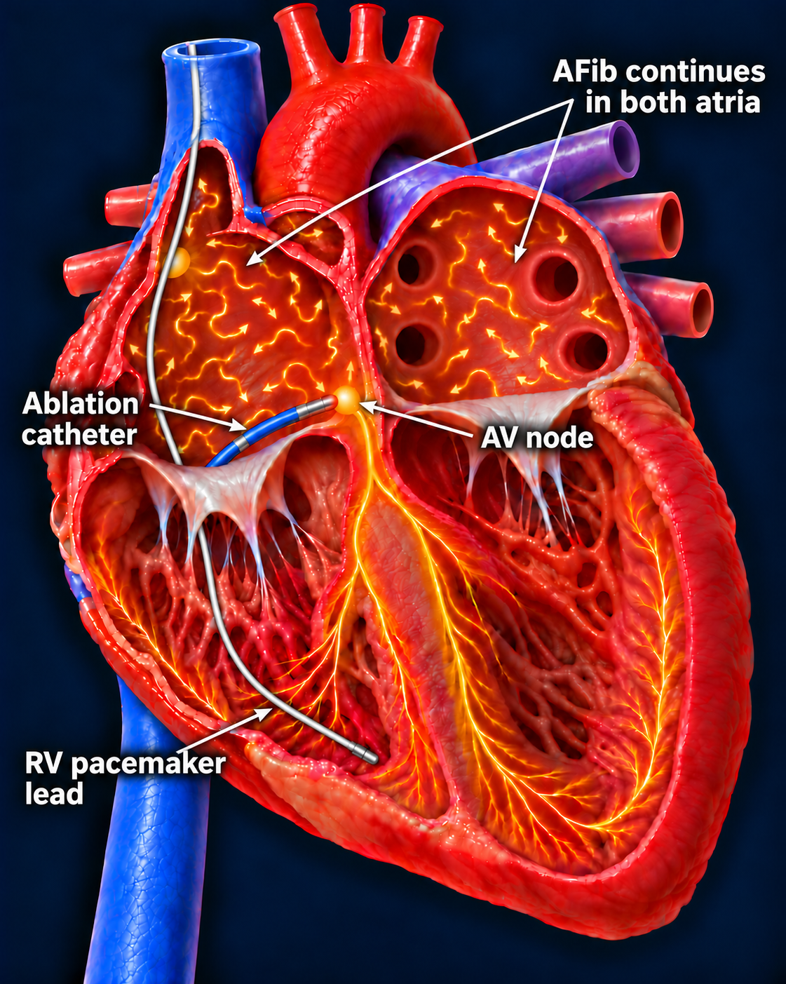
This controls the rate—it does not cure AFib
AFib remains in the upper chambers, and stroke risk remains. Continue blood thinners prescribed for stroke prevention unless your care team changes that plan.
Pillar 3: Managing the heart rhythm
Rhythm management aims to restore a normal heartbeat and reduce AFib episodes. Options include cardioversion, rhythm-control medications, and catheter ablation.
Option 1: Electrical cardioversion — resetting the rhythm
Cardioversion uses a brief, controlled electrical shock delivered through pads on your chest and back. You receive sedation so you are asleep and comfortable during the procedure.
The shock interrupts the disorganized electrical activity and can allow your heart’s normal electrical system to take over again.
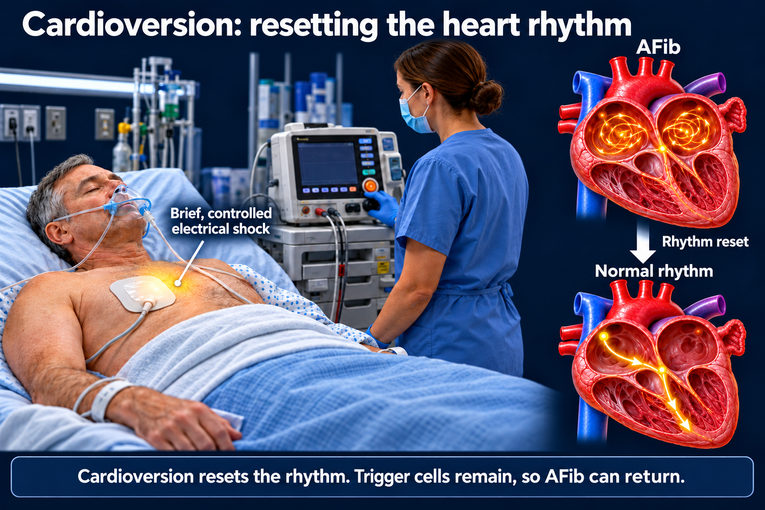
Cardioversion resets the rhythm, but it does not remove or electrically isolate the trigger cells that can start AFib. Those cells remain, so AFib can return—even after a successful cardioversion. Medications or catheter ablation may be recommended to help maintain normal rhythm.
Your care team also plans stroke protection before and after cardioversion. Returning to normal rhythm does not automatically mean you can stop your blood thinner.
Option 2: Rhythm-control medications — helping maintain a normal rhythm
Rhythm-control medications, also called antiarrhythmic medications, help your heart stay in a normal rhythm. They work by changing electrical activity in the heart to reduce how often AFib occurs.
These medications do not cure AFib. Their goal is to suppress it. AFib can still return, even when you take your medication correctly.
Taking your medication as prescribed
These medicines work best when you follow the prescribed schedule. For medications taken regularly, missed doses can reduce their ability to prevent AFib episodes. Do not stop, change the dose, or take extra doses without instructions from your care team.
Side effects and monitoring
Each medication has its own possible side effects. Your cardiologist chooses a medication based on your heart health, kidney function, other medications, and individual needs.
Some medications require regular blood draws to check kidney function, electrolyte levels, or thyroid and liver function. You may also need EKGs to check how the medication affects your heart’s electrical system. Certain medications must be started in the hospital so your rhythm can be monitored closely.
Tell your care team about side effects so they can adjust your treatment safely.
Keeping your heart in normal rhythm does not automatically mean you can stop your blood thinner. Stroke prevention remains a separate part of your AFib treatment.
Option 3: Catheter ablation — targeting AFib at its source
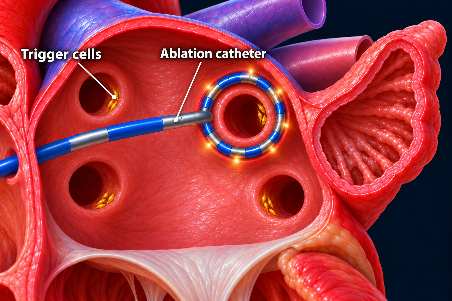
AFib often starts when tiny groups of trigger cells near the openings of the pulmonary veins send abnormal electrical signals into the left atrium. These veins bring blood from the lungs back to the heart.
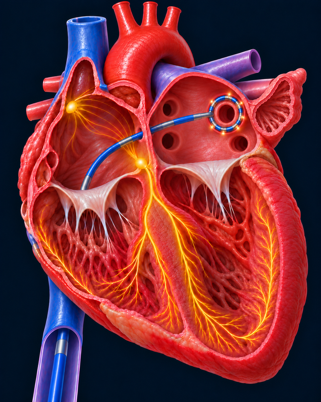
During ablation, your electrophysiologist guides a thin, flexible catheter through a blood vessel into your heart. The catheter delivers energy to disable targeted heart muscle cells and create an electrical barrier around the pulmonary veins. This is called pulmonary vein isolation, or PVI.
The barrier prevents abnormal signals inside the veins from reaching the rest of the atrium and starting AFib. The veins remain open, so blood can still flow normally. Ablation can use heat, freezing, or short electrical pulses called pulsed field ablation.
1. How the catheter reaches the left atrium
2. How ablation isolates the trigger cells
Why choose ablation?
Catheter ablation is generally the most effective of these options for long-term AFib suppression. Cardioversion resets the rhythm, and medications suppress abnormal electrical activity. Ablation directly targets the pathways that allow trigger signals to start AFib.
Compared with rhythm-control medications, ablation generally provides better control of AFib episodes and symptoms. Outcomes are often better when treatment occurs earlier.
Can AFib return?
Ablation is not a guaranteed cure. Electrical connections can recover, or AFib can develop from other areas. Some patients need another procedure or continued medication.
Your stroke-prevention plan remains separate: successful ablation does not automatically mean you can stop your blood thinner.
Preparing for your ablation?
Learn about procedure day, recovery, and caring for your groin puncture sites at home.
What to expect on my procedure dateUpdated October 2026. Educational information; your own care plan may differ.