What should I do now?
Call 911
Call 911 for chest pressure, severe trouble breathing, fainting during a racing heartbeat, or stroke signs such as face drooping, arm weakness, or trouble speaking. Do not drive yourself.
Contact your care team
Contact your care team for new or longer episodes, worsening fatigue, reduced activity, medication side effects, or bleeding concerns.
Keep your care plan
Keep your treatment and follow-up plan. Bring questions, readings, and your medication list.
01 / START WITH THE BASICS
1. Understanding AFib: What is happening in your heart?
First, how does a normal heartbeat work?
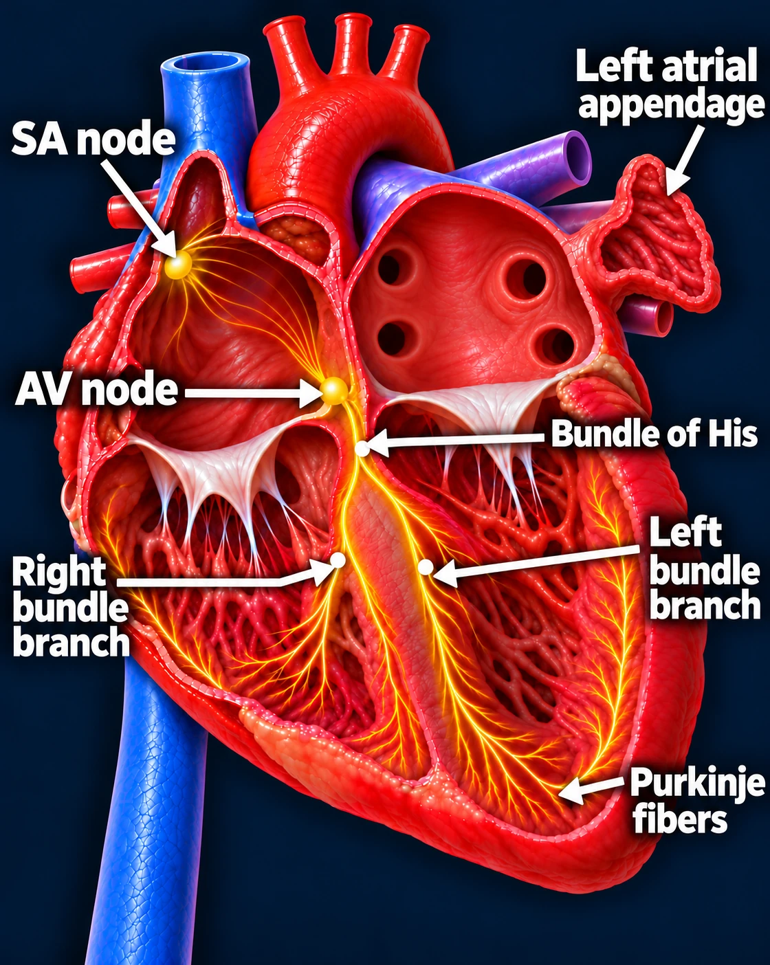
Your heart has an electrical system that coordinates each heartbeat. A normal rhythm begins in the SA node, the heart’s natural pacemaker in the right upper chamber.
Follow one normal heartbeat
SA node → Atria → AV node → Ventricles
- The SA node starts the signal. The heart’s natural pacemaker in the right upper chamber begins each normal sinus beat.
- The signal spreads through both atria. The upper chambers contract, moving blood into the ventricles.
- The AV node briefly delays the signal. This pause gives the ventricles time to finish filling.
- The signal activates the ventricles. It travels through the bundle of His, right and left bundle branches, and Purkinje fibers. The lower chambers contract, pumping blood to the lungs and body.
The atria contract first; the ventricles follow. This organized sequence produces a coordinated heartbeat.
Sinus rhythm
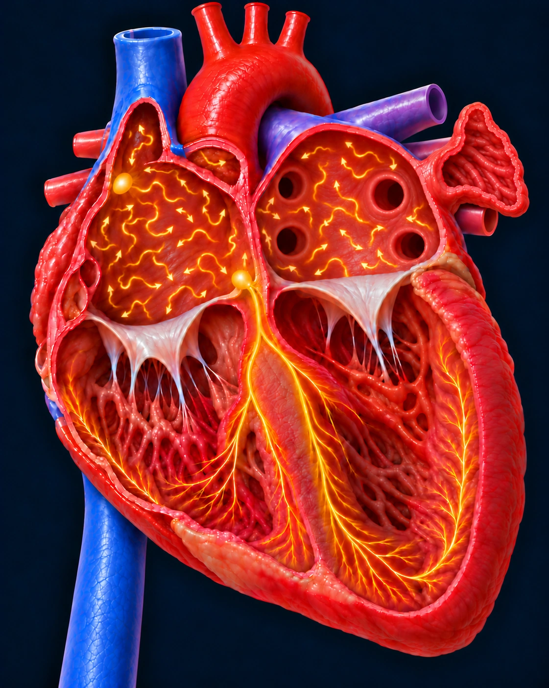
In atrial fibrillation (AFib), these electrical signals become disorganized. Instead of squeezing normally, the atria quiver. This causes an irregular heartbeat that may be fast, even when you do not feel symptoms.
Atrial fibrillation
How does AFib start?
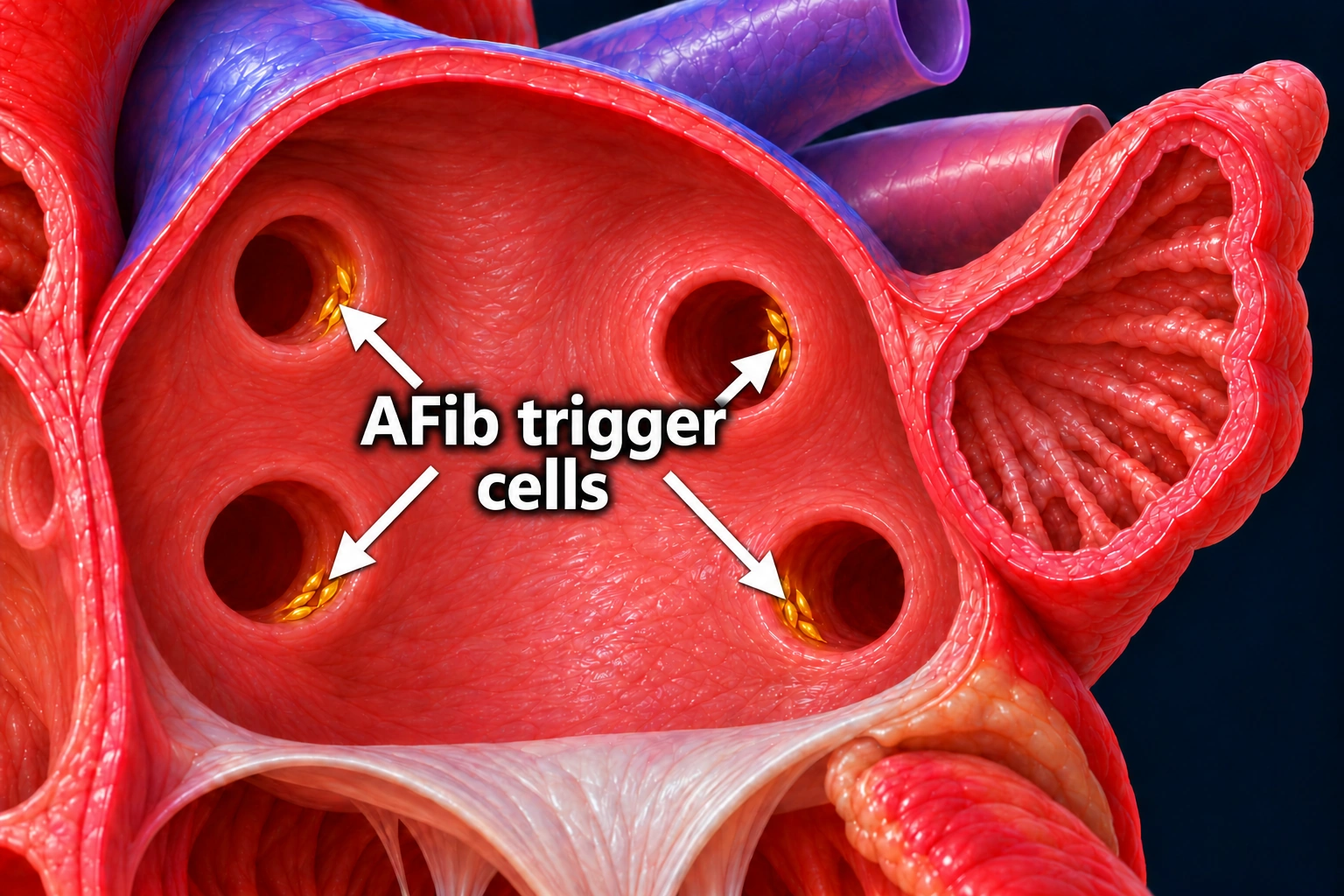
AFib often starts when tiny groups of heart muscle cells near the openings of the pulmonary veins send out extra electrical signals. These veins carry blood from the lungs back into the heart’s upper left chamber, called the left atrium.
We call these cells “trigger cells” because their signals can interrupt the normal heartbeat and trigger an episode of AFib. They are found in small sleeves of heart muscle that extend into the veins near their openings. The extra signals can spread through the atria and start the disorganized electrical activity shown above.
How AFib can lead to a stroke
Because the atria are not squeezing effectively, blood can pool in a small pouch attached to the upper left chamber called the left atrial appendage. When blood sits in this pouch, clots can form. A clot can then break loose, be pumped out of the heart, and travel to the brain. If it blocks an artery supplying the brain, it can cause a stroke.
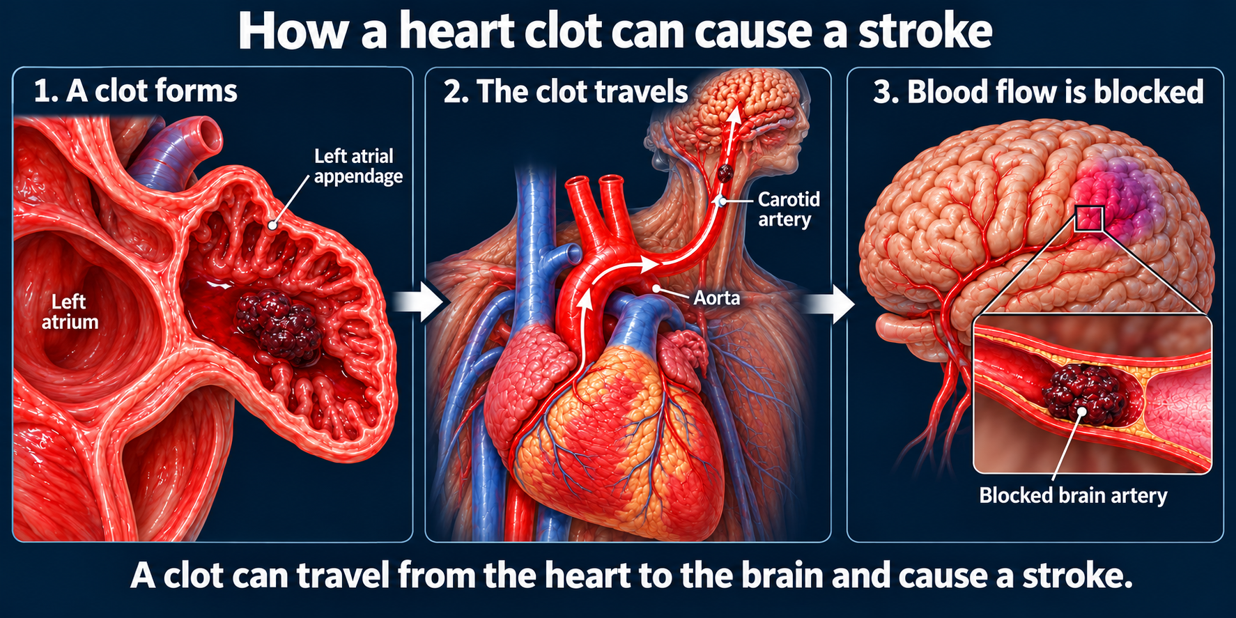
Untreated AFib is associated with about a fivefold increase in stroke risk compared with people without AFib. Your individual risk depends on factors such as your age and other medical conditions. This is why preventing blood clots and stroke is a central part of AFib treatment—even if you feel well.
AFib can progress over time
AFib is a long-term condition with no guaranteed permanent cure. Treatment can provide excellent control—and sometimes years without episodes—but AFib can return.
AFib is also a progressive disease: episodes may become more frequent, last longer, and eventually become continuous. This does not happen to everyone, but longer-lasting AFib can become harder to control.
Why early treatment matters
The goal of rhythm treatment is to suppress AFib, meaning reduce how often it happens and how long it lasts, or maintain a normal rhythm. Your overall treatment plan also addresses symptoms, heart rate, stroke prevention, and conditions that contribute to AFib.
Addressing AFib early gives us an opportunity to improve symptoms and limit progression. In appropriate patients, early treatment to restore or maintain a normal rhythm can also reduce serious cardiovascular complications.
Why AFib should not be ignored
Poorly controlled AFib can lead to repeated emergency visits or hospital stays and can cause or worsen heart failure. AFib also increases stroke risk and is associated with a higher risk of dementia, although researchers are still studying that relationship.
Some patients with a persistently fast heartbeat that cannot be controlled with other treatments may eventually need a pacemaker together with a procedure called AV node ablation. A pacemaker is not a routine treatment for AFib, and most patients do not need one.
AFib is manageable. Early evaluation and a personalized treatment plan can help protect your heart and keep you feeling well.
Heart illustrations use conceptual electrical overlays. EKG strips are illustrative, not patient recordings; timing and voltage are not calibrated.
Updated October 2026. Educational information; your own care plan may differ.