AFIB · THE SIMPLE EXPLANATION
AFib, made clear.
Start with three things: your heart rhythm, stroke prevention, and how ablation works.
01 · YOUR HEART RHYTHM
What is AFib?
An electrical problem in the upper chambers of the heart.
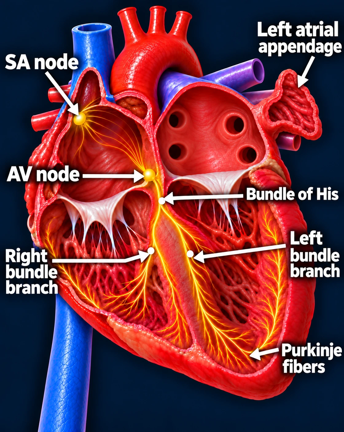
What changes in AFib?
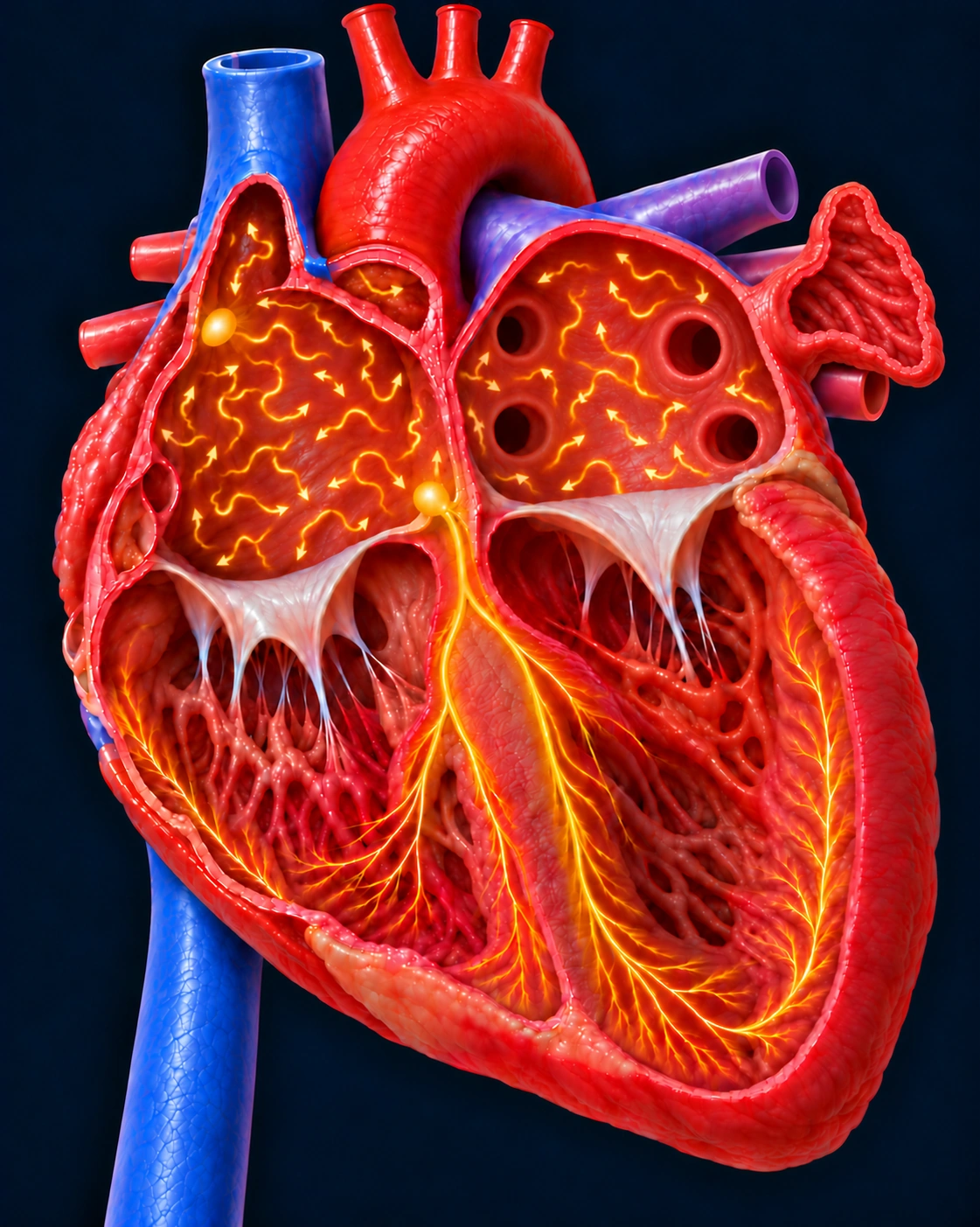
The top chambers (atria) are caught in an electrical storm called fibrillation. Instead of pumping blood forward normally, they quiver.
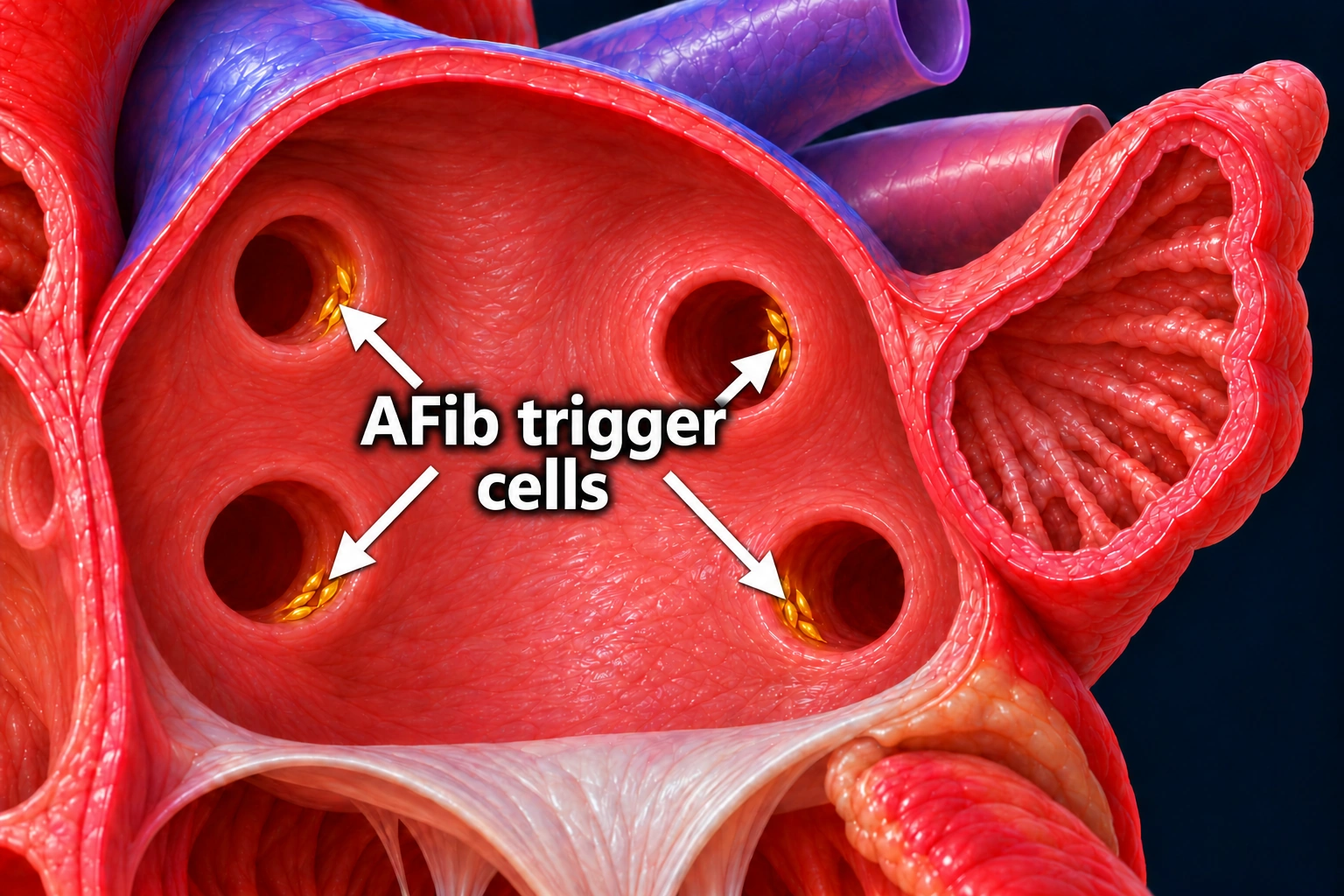
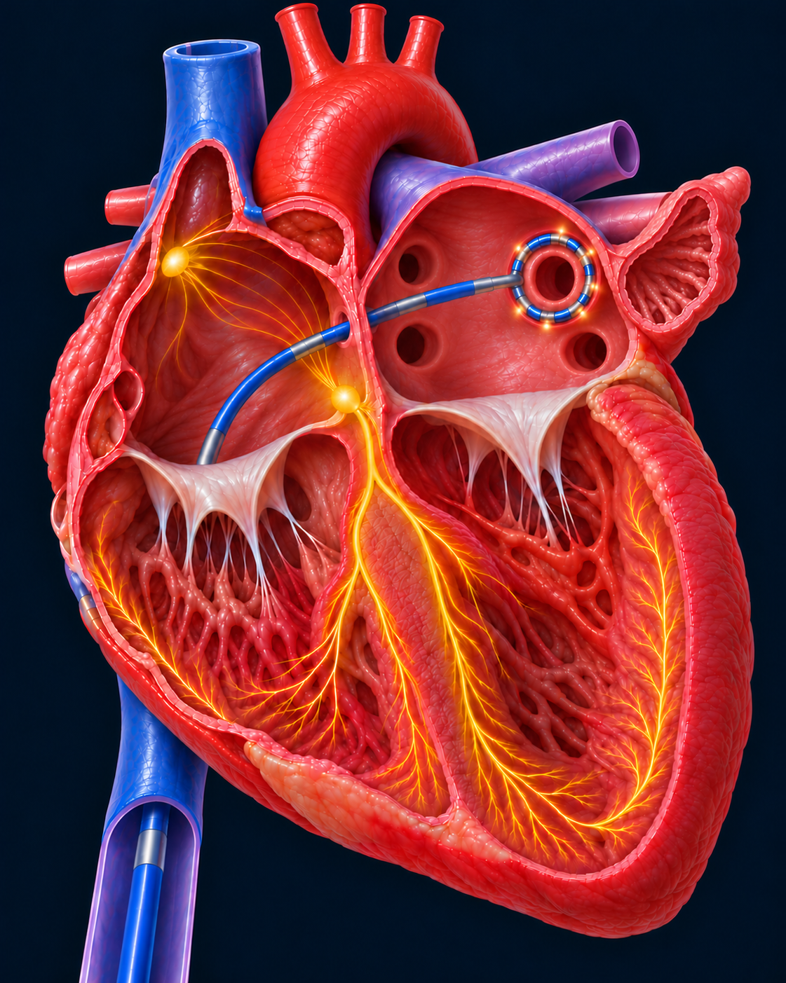
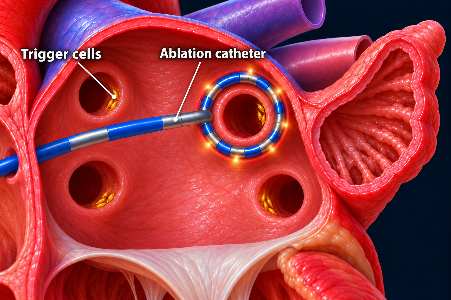
AFib can start when trigger cells in the pulmonary veins send out rapid electrical signals. These veins connect to the left atrium.
You may feel palpitations, tiredness, or shortness of breath—or have no symptoms.
02 · STROKE PREVENTION
Why does AFib increase stroke risk?
Blood thinners lower the chance of clot formation.
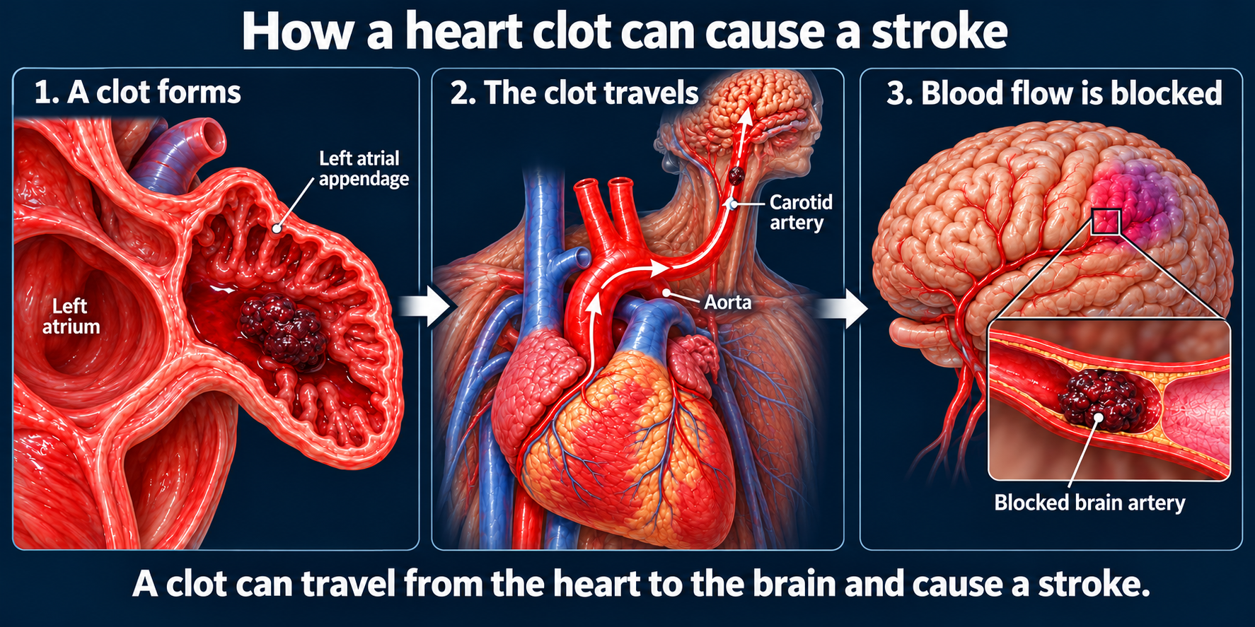 Open full-size stroke illustration →
When the atria do not squeeze normally, blood may pool in a small pouch called the left atrial appendage.
A clot can form, travel to the brain, and cause a stroke.
Blood thinners reduce this risk, but do not eliminate it.
03 · TREATING THE RHYTHM
How does AFib ablation work?
Pulmonary vein isolation (PVI) blocks electrical triggers.
 Open full-size image →
Ablation creates an electrical barrier around the pulmonary veins.
Triggers inside the veins can no longer reach the atrium if isolation remains intact.
Ablation treats the heart rhythm. Blood thinners may still be needed afterward.