What should I do now?
Call 911
Call 911 if racing occurs with chest pressure, severe trouble breathing, fainting, or severe weakness or dizziness. Do not drive yourself.
Contact your care team
Contact your care team if episodes are new, more frequent, longer, or interfere with daily life. Follow your personal plan when an episode will not stop.
Keep your care plan
Keep your treatment and follow-up plan. Bring questions, readings, and your medication list.
03 / DIAGNOSIS
How is SVT diagnosed?
The key is to record your heart’s electrical activity—ideally while the fast rhythm is happening.
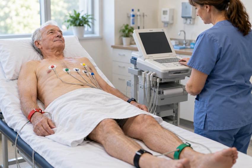
An EKG during an episode
An electrocardiogram, also called an ECG or EKG, uses small electrodes on your chest and limbs to record electrical signals.
The recording helps your clinician distinguish SVT from other causes of a fast heartbeat. It may also provide clues about which type of SVT you have.
Normal rhythm compared with SVT
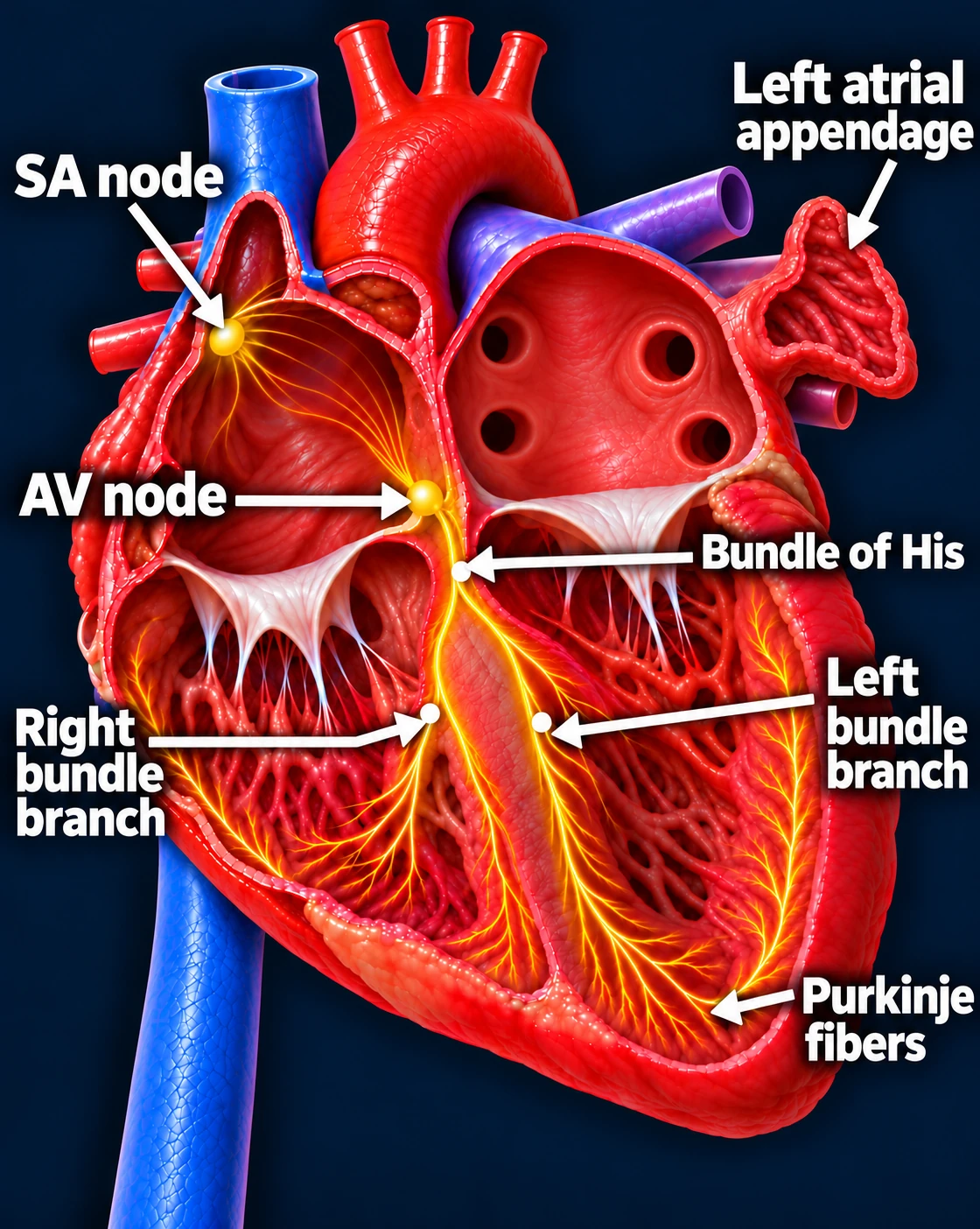
Follow one normal heartbeat
SA node → Atria → AV node → Ventricles
- The SA node starts the signal. The heart’s natural pacemaker in the right upper chamber begins each normal sinus beat.
- The signal spreads through both atria. The upper chambers contract, moving blood into the ventricles.
- The AV node briefly delays the signal. This pause gives the ventricles time to finish filling.
- The signal activates the ventricles. It travels through the bundle of His, right and left bundle branches, and Purkinje fibers. The lower chambers contract, pumping blood to the lungs and body.
The atria contract first; the ventricles follow. This organized sequence produces a coordinated heartbeat.
Normal sinus rhythm
SVT example: AVNRT
Why can my office EKG look normal?
SVT may stop before your appointment. A normal EKG between episodes does not rule it out.
Your clinician may recommend a Holter, patch, or event monitor to capture your rhythm during daily activities. For very infrequent episodes, an implanted loop recorder may sometimes be considered.
Can my smartwatch help?
A device that records an actual EKG may provide useful information. Save the tracing and share it with your care team.
A heart-rate number alone cannot identify the rhythm. A standard blood pressure cuff or finger pulse oximeter can show a fast pulse, but it cannot establish an SVT diagnosis.
How do we determine the exact type?
Sometimes an electrophysiology study, or EP study, is needed. An EP guides thin catheters into the heart to record signals and test the electrical pathways.
This can distinguish AVNRT, AVRT, and atrial tachycardia. When appropriate and agreed upon beforehand, ablation can be performed during the same procedure.
The key idea: Symptoms suggest a rhythm problem. Electrical recordings help diagnose it, and an EP study can identify its precise mechanism.
Updated October 2026. Educational information; your own care plan may differ.