What should I do now?
Call 911
Call 911 if racing occurs with chest pressure, severe trouble breathing, fainting, or severe weakness or dizziness. Do not drive yourself.
Contact your care team
Contact your care team if episodes are new, more frequent, longer, or interfere with daily life. Follow your personal plan when an episode will not stop.
Keep your care plan
Keep your treatment and follow-up plan. Bring questions, readings, and your medication list.
01 / UNDERSTAND SVT
What is SVT?
Supraventricular tachycardia, or SVT, is a group of abnormal heart rhythms that make the heart beat faster than it should. “Supraventricular” means coming from the heart’s upper chambers. “Tachycardia” means a fast heart rate.
Many common SVTs cause episodes of a rapid, regular heartbeat that start and stop suddenly—almost like flipping a switch. An episode may occur while you are resting or active.
How does the heart normally beat?
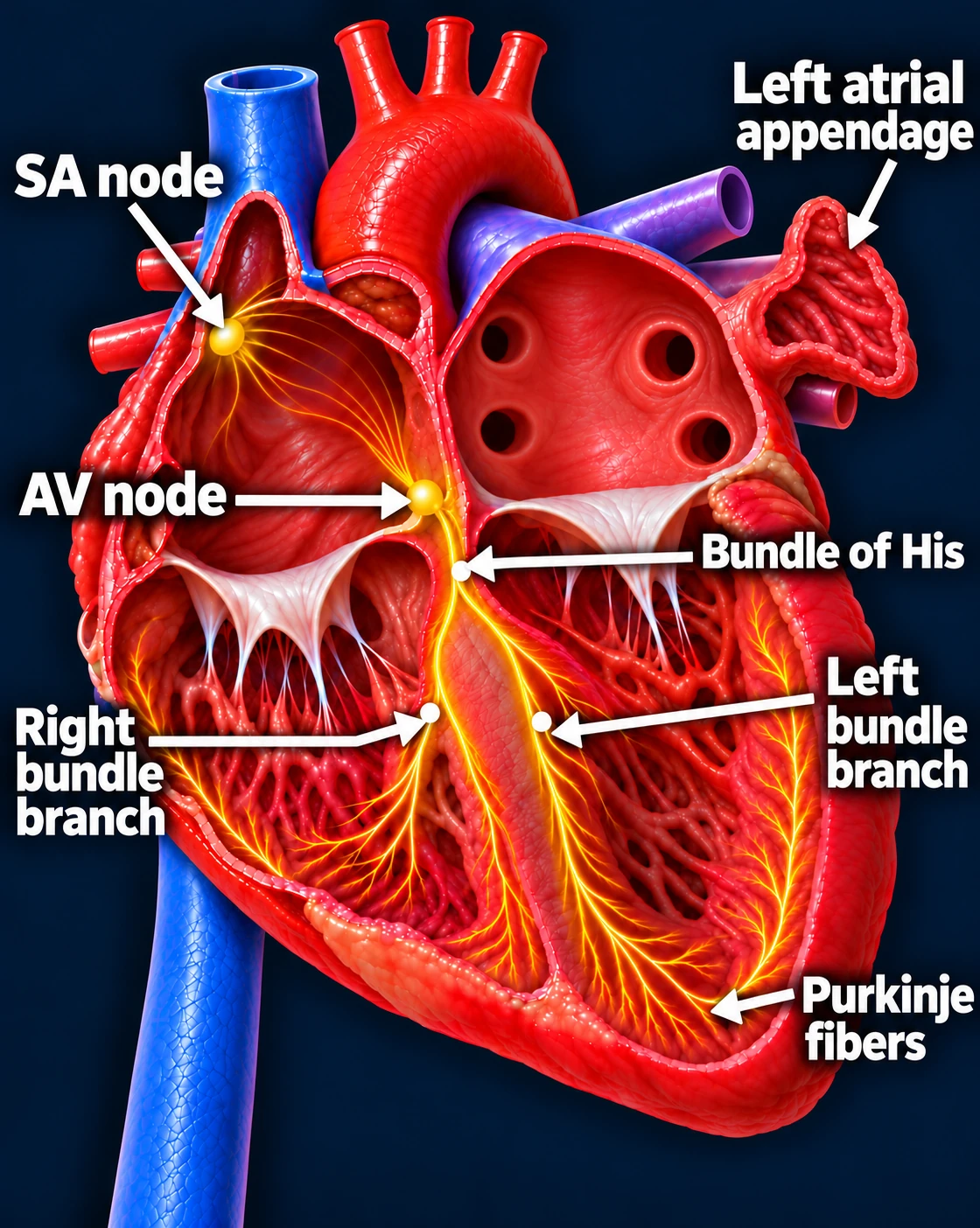
Follow one normal heartbeat
SA node → Atria → AV node → Ventricles
- The SA node starts the signal. The heart’s natural pacemaker in the right upper chamber begins each normal sinus beat.
- The signal spreads through both atria. The upper chambers contract, moving blood into the ventricles.
- The AV node briefly delays the signal. This pause gives the ventricles time to finish filling.
- The signal activates the ventricles. It travels through the bundle of His, right and left bundle branches, and Purkinje fibers. The lower chambers contract, pumping blood to the lungs and body.
The atria contract first; the ventricles follow. This organized sequence produces a coordinated heartbeat.
Sinus rhythm
What changes during SVT?
In common types of SVT, an electrical signal becomes trapped in a repeating loop, or an abnormal electrical source sends signals faster than the sinus node. These signals take over the rhythm and drive the heart too quickly.
SVT is an electrical problem. Having SVT does not necessarily mean you have blocked arteries or a weak heart.
There are multiple types of SVT
There are several types of SVT. Here, we’ll focus on three of the main types: AVNRT, AVRT, and atrial tachycardia. Each involves a different electrical pathway or source. Understanding which type you have helps explain why episodes happen and how they can be treated.
Type 1: AVNRT—a loop near the AV node
AV nodal reentrant tachycardia, or AVNRT, occurs when two electrical pathways in or near the AV node allow a signal to circle repeatedly.
These are often called the fast pathway and the slow pathway. Under the right conditions, a premature heartbeat can start a loop that travels down one pathway and back up the other. Each trip around the loop triggers another heartbeat.
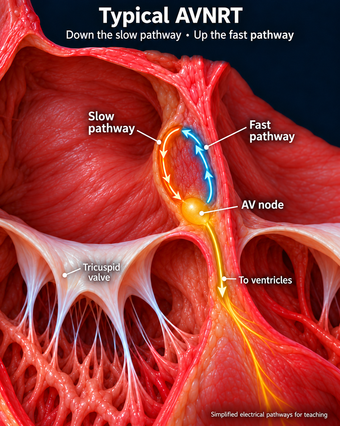
Typical AVNRT travels down the slow pathway (orange) and back up the fast pathway (blue). These functional pathways are enlarged and simplified for teaching.
AVNRT: example EKG
Type 2: AVRT—a loop using an extra connection
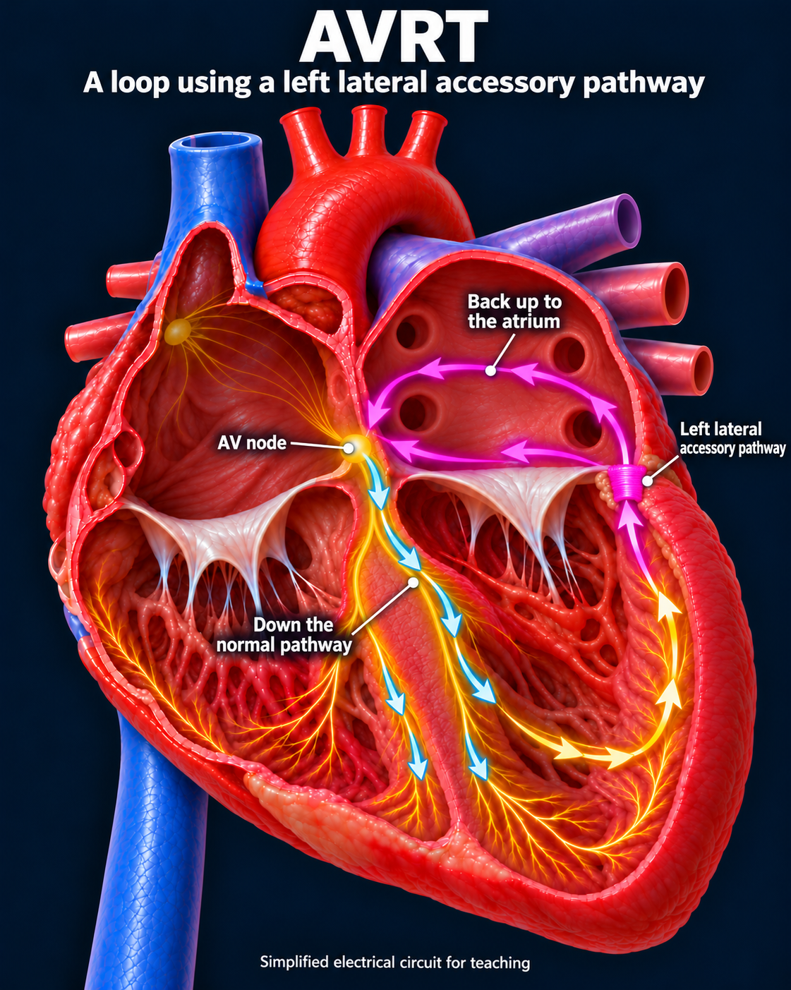
AV reentrant tachycardia, or AVRT, uses an extra electrical connection between the atria and ventricles, called an accessory pathway.
The signal can travel down the normal electrical route and return through the extra connection, creating a repeating loop.
Some accessory pathways are associated with Wolff-Parkinson-White, or WPW. Others do not show up on an EKG taken during normal rhythm.
Orthodromic AVRT: example EKG
Type 3: Atrial tachycardia—an abnormal electrical source
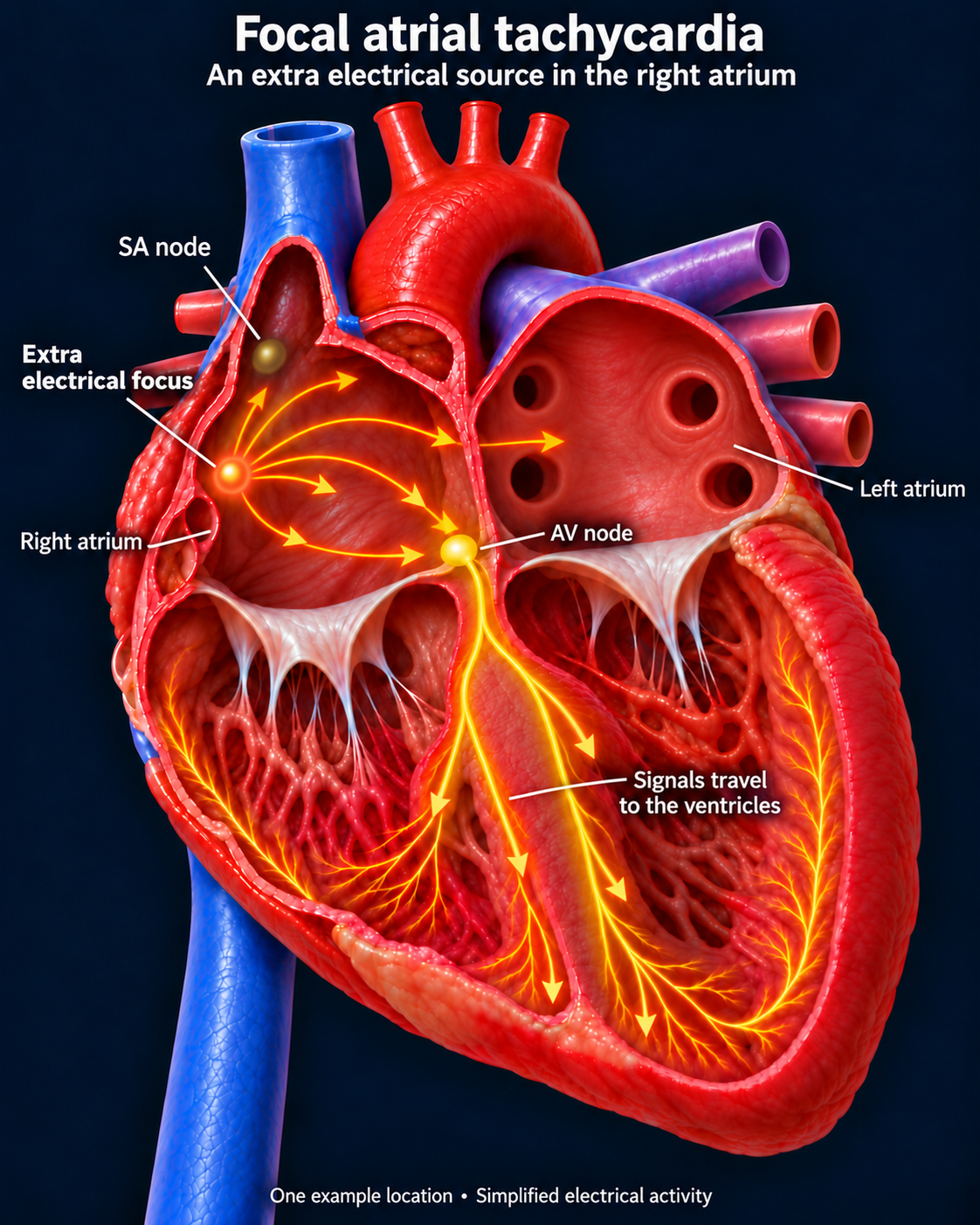
In focal atrial tachycardia, a small area in an atrium sends electrical signals faster than the sinus node. This area temporarily takes over as the rhythm’s driver.
Unlike AVNRT or AVRT, this rhythm does not require a loop through the AV node to keep going.
Atrial tachycardia: example EKG
Why does the type matter?
Different SVTs require different treatment targets. An electrophysiologist, or EP, identifies the electrical pathway or source responsible for your rhythm and discusses the best treatment options.
Many common SVTs can be treated very effectively with catheter ablation, which targets the tissue sustaining the abnormal rhythm.
The key idea: SVT describes several fast electrical rhythms. Understanding which type you have helps explain why it happens—and how we can treat it.
Do I need a blood thinner for SVT?
AVNRT, AVRT, and focal atrial tachycardia do not routinely require blood thinners for stroke prevention. They do not carry the same established clot-related stroke risk as AFib or atrial flutter.
You may still need a blood thinner for another condition. Do not stop a prescribed blood thinner without discussing it with your care team.
Updated October 2026. Educational information; your own care plan may differ.