What should I do now?
Call 911
Call 911 for chest pressure, severe trouble breathing, fainting during a racing heartbeat, or stroke signs. Do not drive yourself.
Contact your care team
Contact your care team for recurrent racing, worsening fatigue, reduced activity, or medication and bleeding concerns.
Keep your care plan
Keep your treatment and follow-up plan. Bring questions, readings, and your medication list.
01 / UNDERSTAND THE RHYTHM
1. Understanding atrial flutter
First, how does a normal heartbeat work?
Your heart has an electrical system that coordinates each heartbeat. A normal rhythm begins in the SA node, the heart’s natural pacemaker in the right upper chamber.
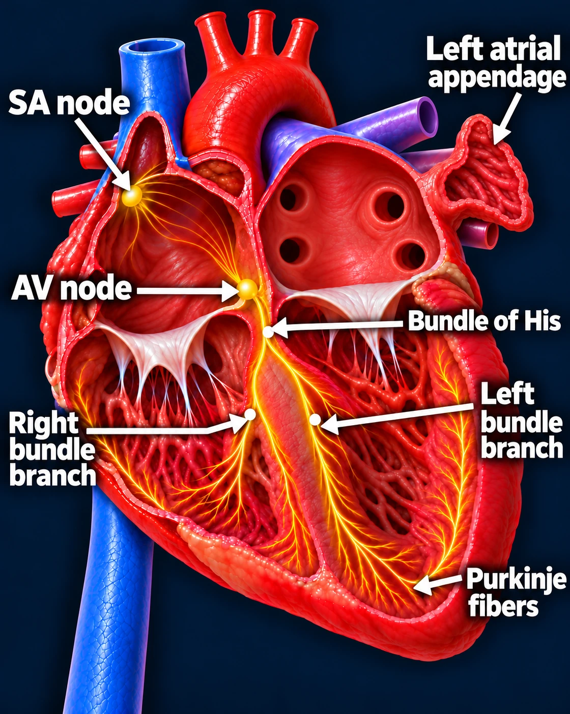
Follow one normal heartbeat
SA node → Atria → AV node → Ventricles
- The SA node starts the signal. The heart’s natural pacemaker in the right upper chamber begins each normal sinus beat.
- The signal spreads through both atria. The upper chambers contract, moving blood into the ventricles.
- The AV node briefly delays the signal. This pause gives the ventricles time to finish filling.
- The signal activates the ventricles. It travels through the bundle of His, right and left bundle branches, and Purkinje fibers. The lower chambers contract, pumping blood to the lungs and body.
The atria contract first; the ventricles follow. This organized sequence produces a coordinated heartbeat.
Sinus rhythm
What changes during atrial flutter?
In atrial flutter, an electrical signal becomes trapped in a repeating loop in the upper chambers. Each trip around the loop activates the atria again, making them beat much faster than normal.
The repeating circuit takes over the rhythm from the SA node. The atria often activate around 250–350 times per minute.
Typical flutter: a circuit in the right atrium
In the most common form, called typical atrial flutter, the signal circles around the tricuspid valve in the right atrium.
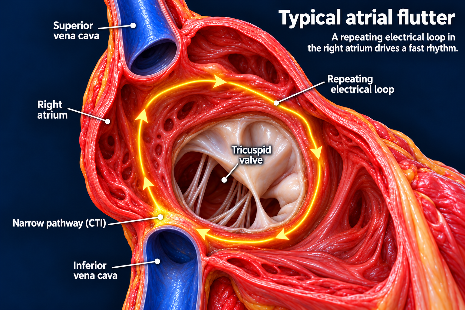
The circuit passes through a narrow bridge of tissue between the tricuspid valve and the inferior vena cava—the large vein bringing blood back from the lower body. This bridge is called the cavotricuspid isthmus, or CTI.
Normal rhythm: Each heartbeat begins with a new signal from the SA node.
Atrial flutter: A signal keeps circling through the same pathway, activating the atria repeatedly.
This narrow CTI pathway is important because an ablation can interrupt the circuit there.
Why isn’t the pulse always 300?
The AV node usually allows only some flutter signals to reach the ventricles.
For example, if the atria activate 300 times per minute and every second signal reaches the ventricles, the pulse is approximately 150 beats per minute. This is called 2:1 conduction.
Other ratios can occur, and conduction can change over time. The pulse may therefore be regular or irregular, fast or relatively controlled.
The rapid atrial rhythm disrupts the normal timing between the upper and lower chambers. A persistently fast ventricular rate can also strain and weaken the heart.
Example EKG: typical flutter
Classic typical flutter can produce small, repeating “sawtooth” flutter waves. The tall QRS spikes represent ventricular beats. This illustrative example shows 2:1 conduction: atrial rate about 300, ventricular rate about 150 beats per minute.
Atypical flutter: a different circuit
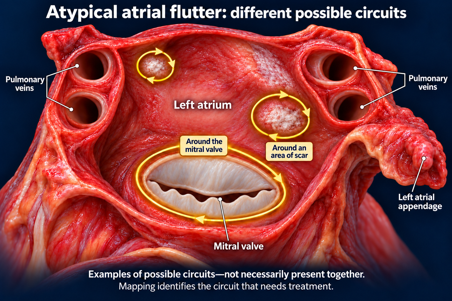
Atypical flutter follows a pathway outside the typical CTI circuit. It can occur in either atrium, sometimes around scar tissue from heart disease, previous surgery, or an earlier ablation.
An electrophysiologist uses electrical mapping to identify the circuit and determine how best to treat it.
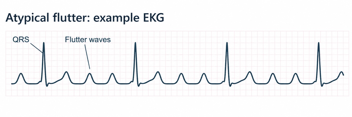
Example EKG: atypical flutter
 Open larger image (new tab)
This example shows several rounded atrial waves between the QRS complexes, without a classic sawtooth pattern. Atypical flutter does not have one fixed EKG appearance.
EKG strips are illustrations, not patient recordings. Flutter-wave appearance varies, and the EKG alone may not identify the exact circuit or reliably distinguish typical from atypical flutter.
Flutter and AFib: related, but different
Flutter usually involves an organized, repeating electrical circuit. AFib involves disorganized electrical activity. A person can have both conditions, and treating flutter does not necessarily prevent AFib.
Why can flutter increase stroke risk?
When the atria beat rapidly, they may not empty effectively. Blood can stagnate, particularly in the left atrial appendage, allowing a clot to form. If a clot leaves the heart and blocks an artery in the brain, it can cause a stroke.
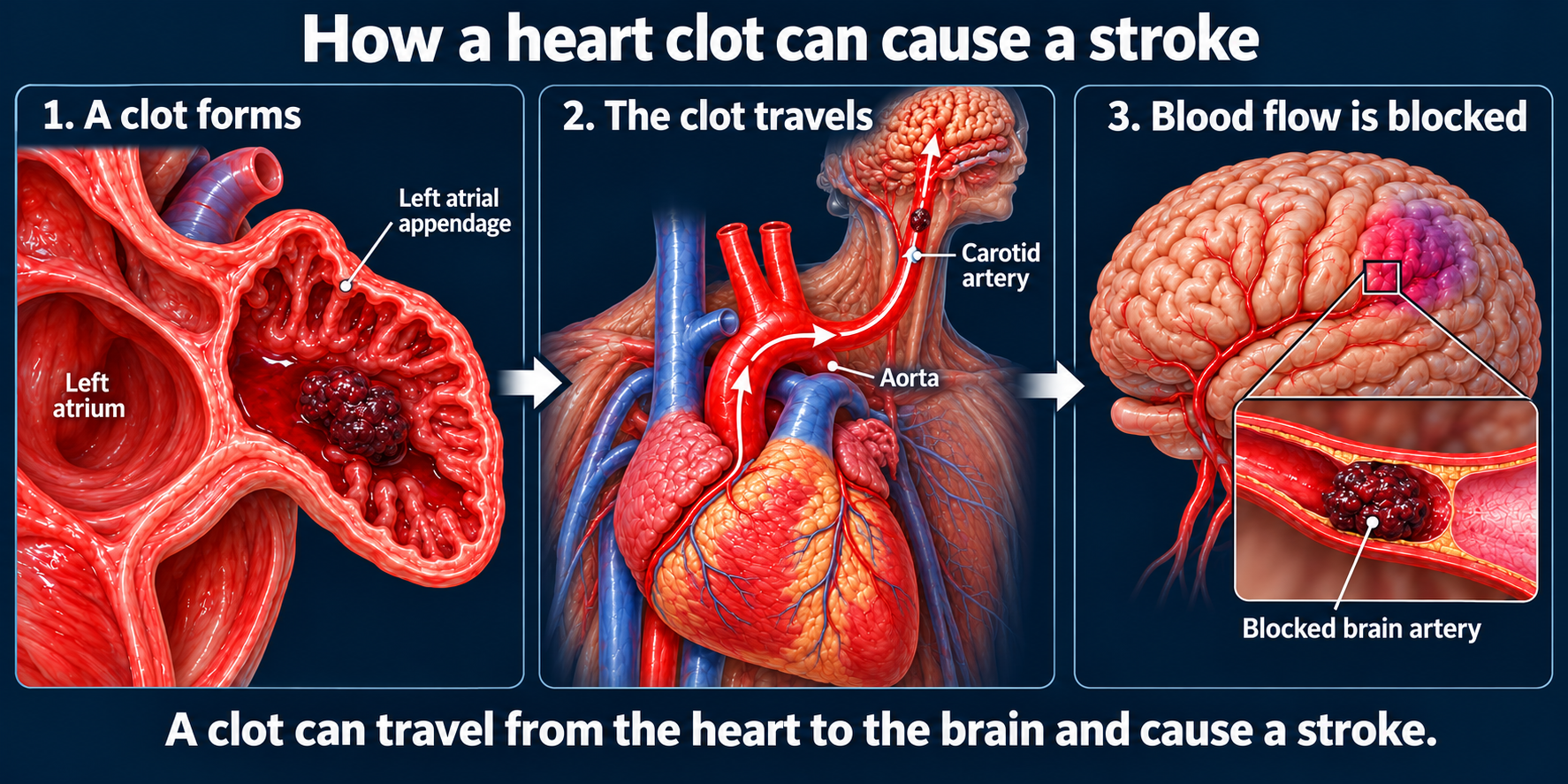
Both rhythms can increase stroke risk. Even when the pulse feels regular or symptoms are mild, your care team needs to assess whether you need a blood thinner.
The goals are to protect against stroke, control the heart rate, and restore a normal rhythm when appropriate.
Heart illustrations use conceptual electrical overlays. EKG strips are illustrative, not patient recordings; timing and voltage are not calibrated.
Updated October 2026. Educational information; your own care plan may differ.