What should I do now?
Call 911
Call 911 for new or unexplained chest pressure that persists or returns, severe trouble breathing, collapse without normal recovery, or sudden face/arm weakness or trouble speaking. Do not drive yourself.
Contact your care team
Contact your care team for persistent or worsening fatigue or a meaningful decline in your usual activity.
Keep your care plan
Record how symptoms affect daily tasks, and review sleep and medications with your clinician.
SECTION 2
What heart problems can cause low energy?
Heart problems can reduce stamina by affecting pumping, filling, blood supply, or rhythm. More than one cause may be present.
Heart failure: weaker pumping or difficulty filling
A weakened heart may struggle to meet the body’s needs during activity. Heart failure can also occur when the heart squeezes normally but does not relax and fill well.
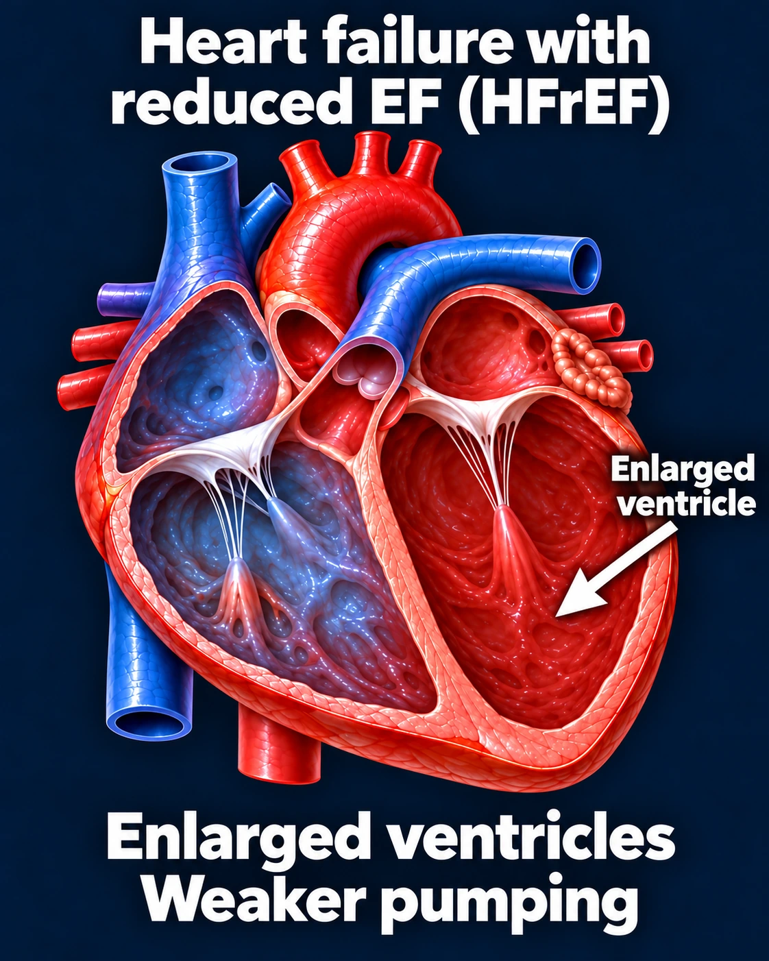
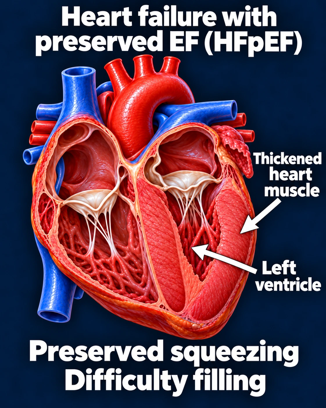
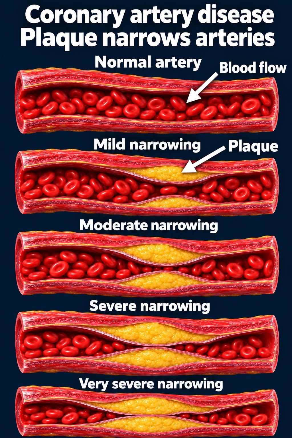
Coronary artery disease: reduced blood supply
Narrowed coronary arteries can limit oxygen-rich blood reaching the heart muscle, particularly during exertion. Some patients notice breathlessness or reduced stamina rather than obvious chest pain.
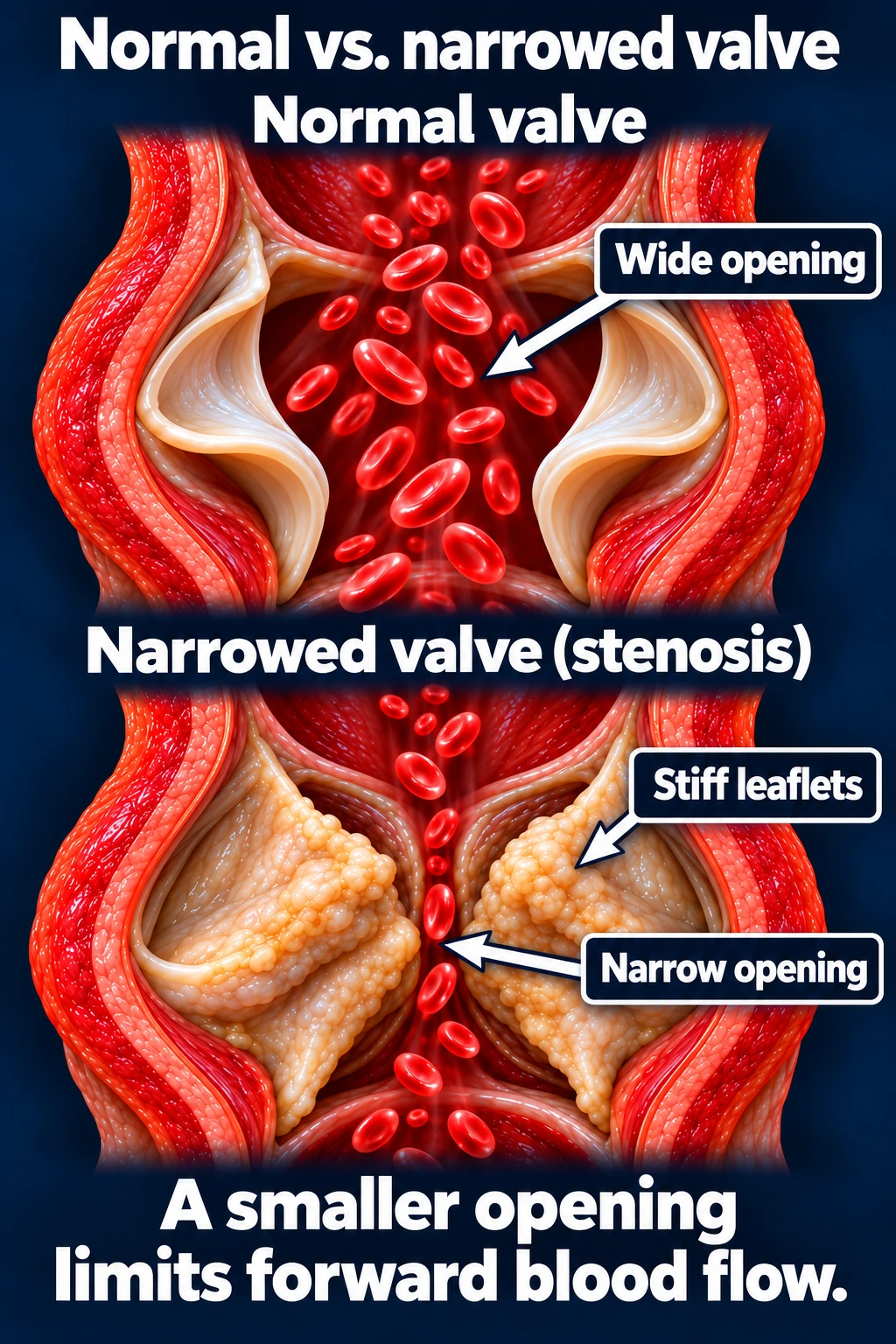
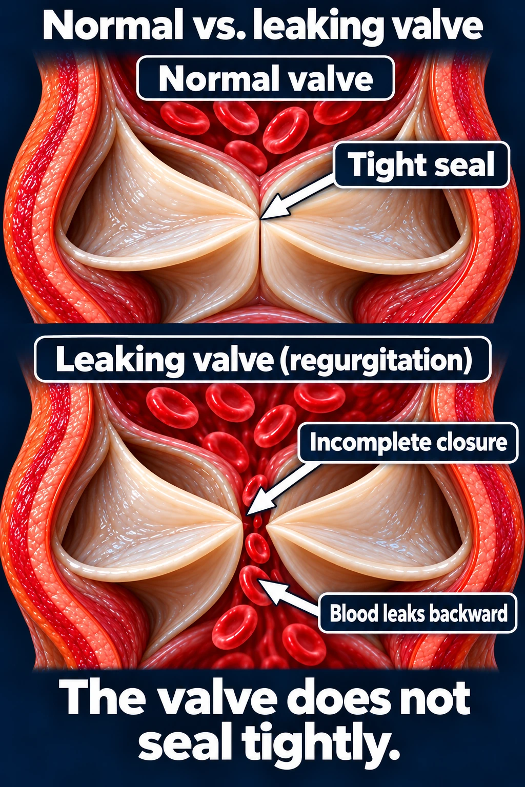
Valve disease: restricted forward flow or a leak
A significantly narrowed valve can obstruct blood flow. A significant leak allows some blood to move backward and adds work for the heart. Either can contribute to fatigue and breathlessness.
Slow heart rhythms and heart block
A very slow pulse, blocked electrical signals, or a pulse that does not rise appropriately during activity can reduce energy. Some people have a naturally slow resting pulse without symptoms.
The normal pacemaker or electrical conduction system can sometimes limit the heart’s response to activity.
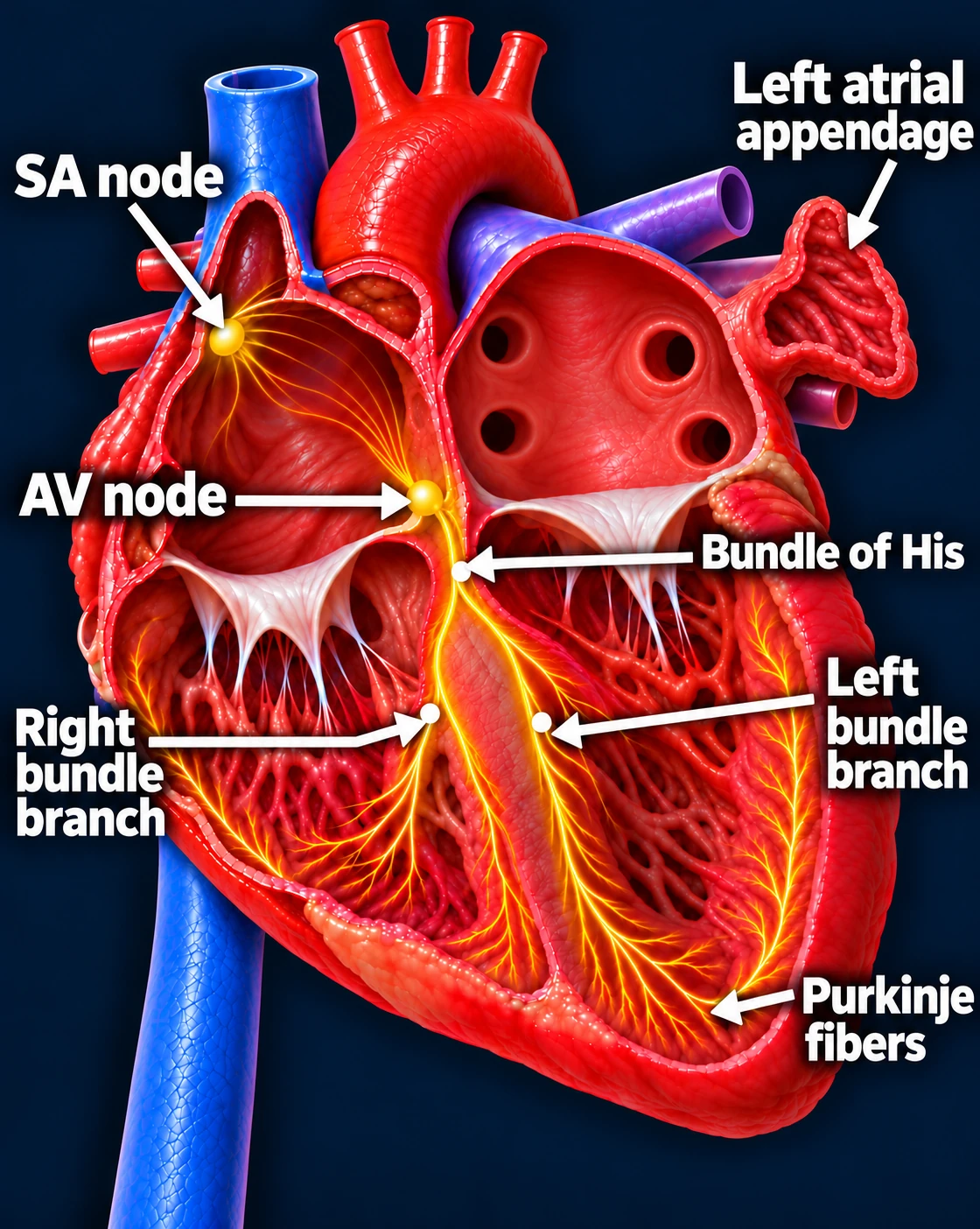
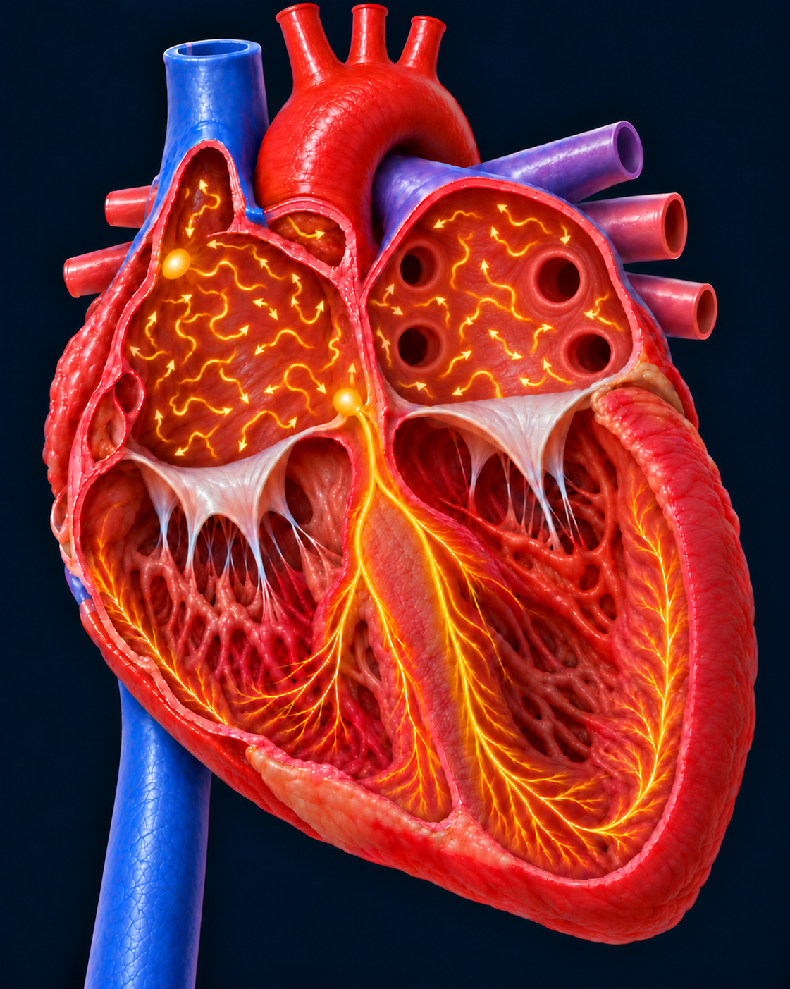
Follow one normal heartbeat
SA node → Atria → AV node → Ventricles
- The SA node starts the signal. The heart’s natural pacemaker in the right upper chamber begins each normal sinus beat.
- The signal spreads through both atria. The upper chambers contract, moving blood into the ventricles.
- The AV node briefly delays the signal. This pause gives the ventricles time to finish filling.
- The signal activates the ventricles. It travels through the bundle of His, right and left bundle branches, and Purkinje fibers. The lower chambers contract, pumping blood to the lungs and body.
The atria contract first; the ventricles follow. This organized sequence produces a coordinated heartbeat.
Normal sinus rhythm — comparison
A very slow heartbeat — sinus bradycardia
The beats are farther apart. Whether a slow rate causes fatigue depends on the patient and the heart’s response to activity.
Complete heart block
Signals from the upper chambers do not reach the lower chambers. A slower backup rhythm runs independently.
Fast or irregular rhythms
AFib, atrial flutter, and SVT can reduce exercise tolerance, especially when the pulse is rapid. They may affect filling and the coordination of the heartbeat.
Atrial fibrillation — irregular rhythm

SVT — a fast, regular rhythm

These EKGs are simplified teaching examples. They do not show whether a particular rhythm is causing your fatigue. Heart failure, coronary disease, and valve disease may be present even with a normal rhythm strip.
Medicines and causes outside the heart
Some heart medicines can contribute to fatigue. Poor sleep or sleep apnea, anemia, thyroid disease, lung disease, infection, depression, and reduced conditioning can also cause low energy. Do not stop prescribed medicines on your own.
Sources: American Heart Association: Heart failure symptoms · Valve disease symptoms · AFib symptoms
Updated October 2026. Educational information; your own care plan may differ. Medical illustrations are simplified examples for teaching.