What should I do now?
Call 911
Call 911 for collapse, fainting with racing, chest pressure, severe trouble breathing, or repeated ICD shocks. If someone is unresponsive and not breathing normally, follow the dispatcher's CPR/AED instructions.
Contact your care team
After one ICD shock with complete recovery, contact your device team promptly and follow your personal plan. Report recurrent racing or new symptoms.
Keep your care plan
Keep your treatment and follow-up plan. Bring questions, readings, and your medication list.
SECTION 4
How are VT and VF treated?
Treatment has three main goals: stop a dangerous episode, reduce the chance of future episodes, and protect against sudden cardiac death.
Your treatment plan depends on the type of rhythm, its cause, your heart’s pumping function, and your overall health. Several treatments may be used together.
1. Stopping a dangerous episode
VT with a pulse: If VT causes low blood pressure, fainting, chest pain, or other signs that the heart is not pumping adequately, an electrical shock may be needed immediately. This is called cardioversion. Some patients who are stable can receive medication through an IV under close monitoring.
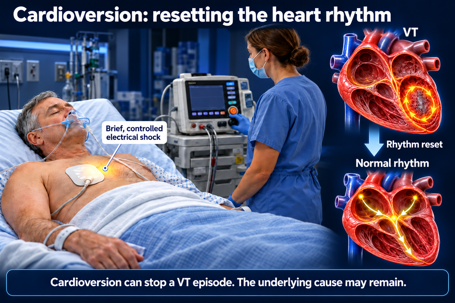
VF or VT without a pulse: These rhythms cause cardiac arrest and require immediate CPR and defibrillation. A defibrillator delivers a shock to try to restore an organized rhythm.
An electrical shock can stop the episode, but it does not necessarily remove the underlying problem that caused it.
2. Treating the underlying cause
Your care team looks for problems that may trigger or worsen ventricular arrhythmias. Treatment may include:
- Correcting low potassium, magnesium, or other electrolyte problems.
- Treating reduced blood flow to the heart or a heart attack.
- Adjusting medications that may contribute to the rhythm.
- Treating heart failure or another heart muscle condition.
- Providing treatment for an inherited electrical disorder.
Treating the cause is an essential part of reducing future risk.
3. An ICD: protection against dangerous rhythms
An implantable cardioverter-defibrillator (ICD) is a device that continuously watches your heart rhythm.
Depending on the device and rhythm, it may deliver:
- Antitachycardia pacing: A short series of fast pacing impulses that can interrupt certain VT episodes.
- An electrical shock: To stop a dangerous rhythm such as VF or VT that cannot be stopped with pacing.
An ICD treats dangerous episodes when they occur. It does not remove scar tissue or prevent every episode from starting. Medications or ablation may still be needed.
Not everyone with a brief run of VT needs an ICD. Your doctor considers your heart function, the cause of the rhythm, previous episodes, and whether a reversible problem was responsible.
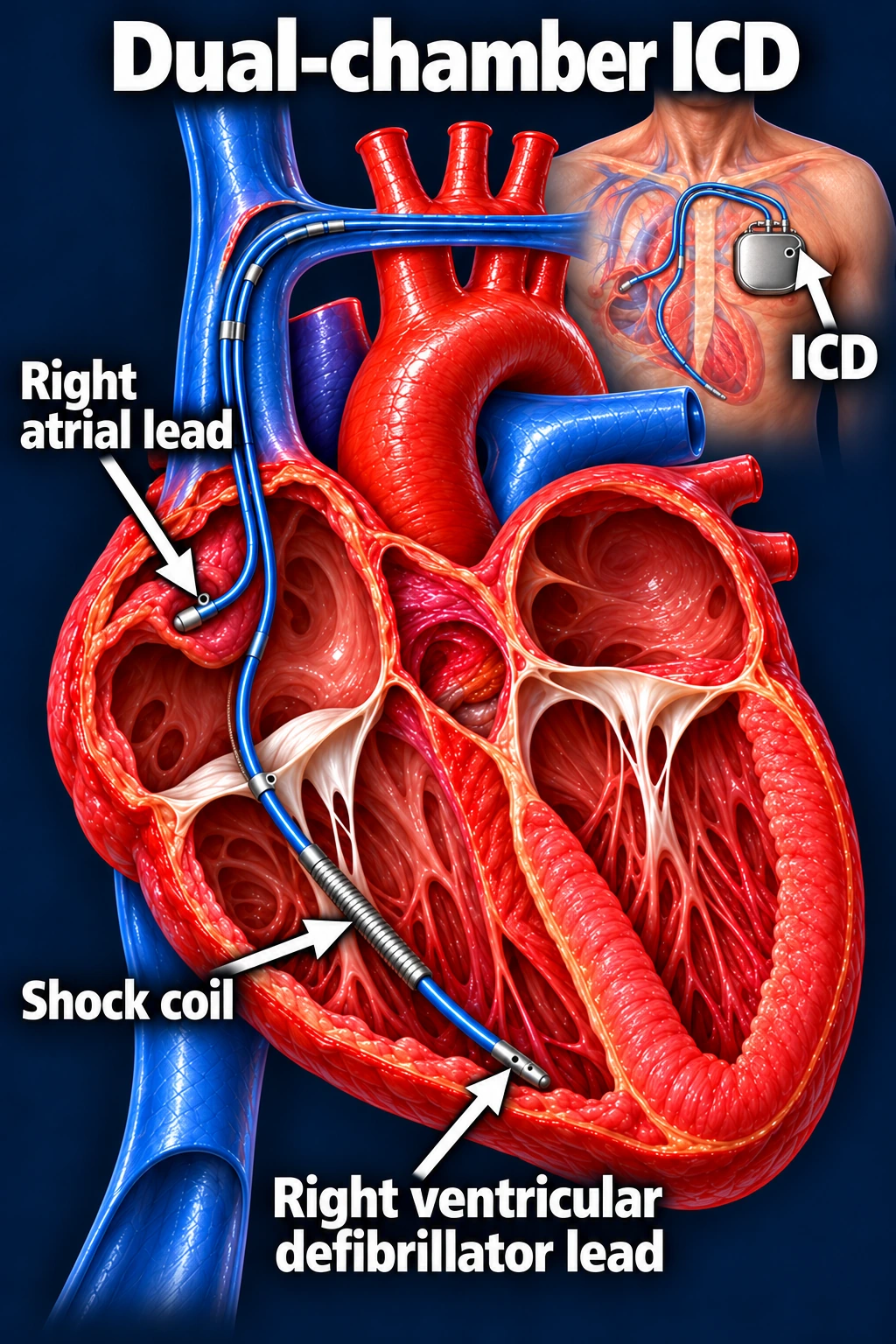
Dual-chamber defibrillator
A dual-chamber ICD has one lead in the right atrium and one in the right ventricle. The ventricular lead includes a shock coil that allows the device to deliver defibrillation therapy.
Biventricular defibrillator (CRT-D)
A CRT-D combines defibrillator protection with pacing that helps the ventricles squeeze together in selected patients with heart failure.
This example has three leads: one in the right atrium, one in the right ventricle, and a third that travels through the coronary sinus to pace the outside of the left ventricle.
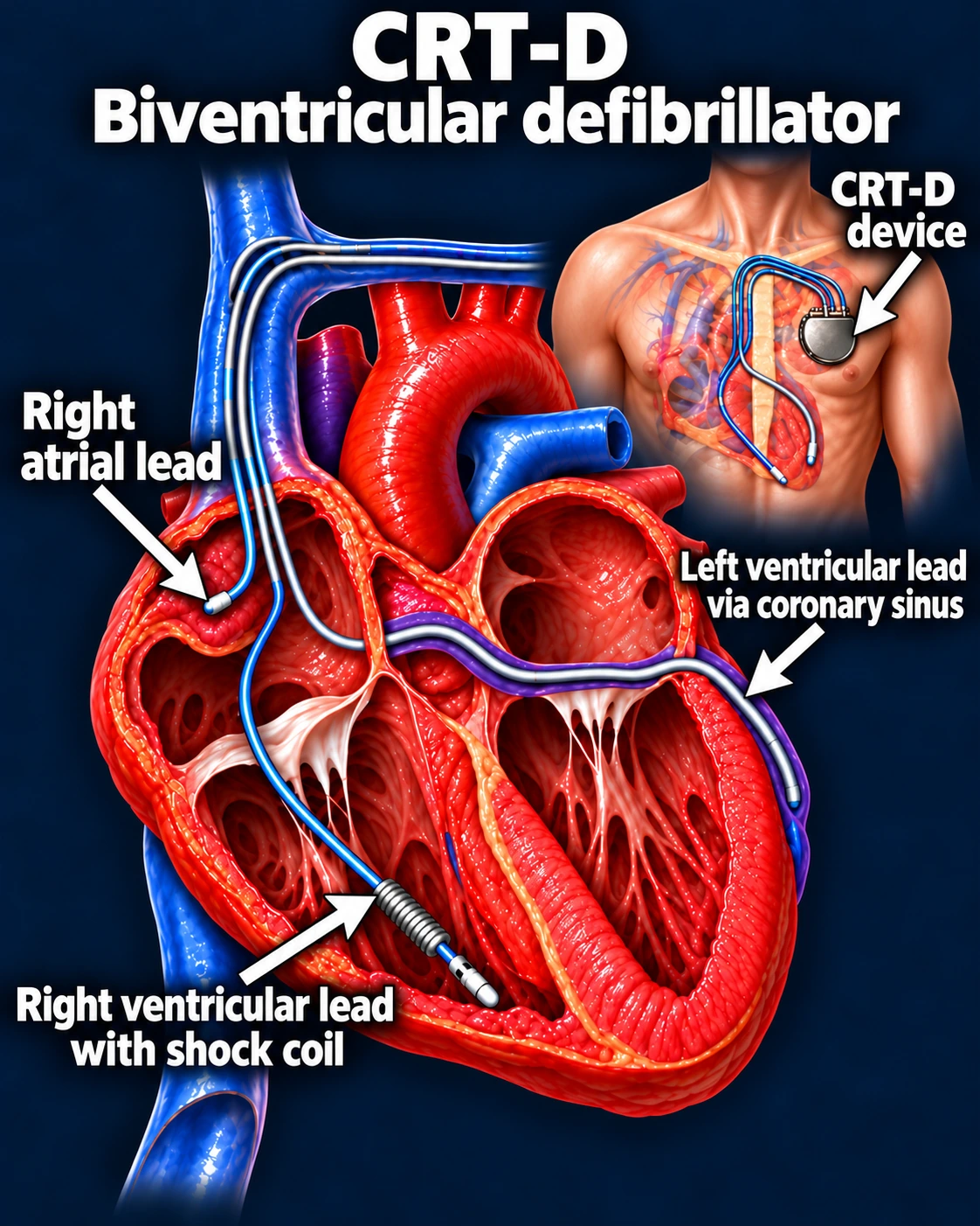
These are two examples of ICD systems. Your physician recommends the type that best fits your rhythm, pacing needs, heart function, anatomy, and overall health.
A wearable defibrillator vest (LifeVest)
Some patients need temporary protection from dangerous VT or VF while their heart function is reassessed or their care team decides on long-term treatment. Your doctor may prescribe a wearable cardioverter-defibrillator, often called a LifeVest.
The vest is worn directly against your skin, underneath clothing, with a portable monitor. It watches your heart rhythm and can automatically deliver a shock for certain life-threatening fast rhythms.
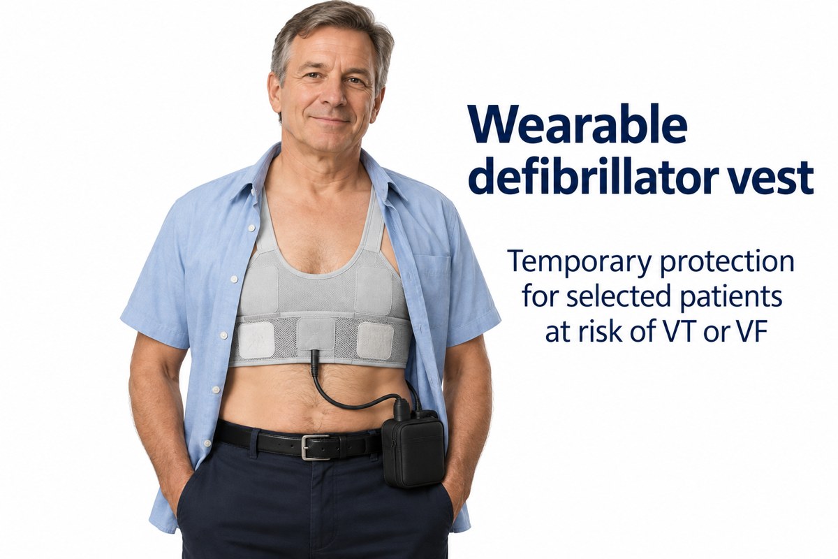
Protection depends on wearing the device as directed. Follow your device team's instructions for wear time, alarms, bathing, and battery changes. The vest treats dangerous episodes; it does not cure the underlying heart condition. Your physician determines whether you need ongoing protection with an implanted ICD.
4. Medications to reduce episodes
Beta-blockers and, in selected patients, antiarrhythmic medications can reduce ventricular arrhythmias.
These medications must be taken as prescribed. Each has potential side effects, and some require follow-up EKGs, blood tests, or other monitoring.
Medication can help suppress episodes, but it does not replace an ICD when an ICD is needed for protection against sudden death.
5. Catheter ablation
During catheter ablation, an electrophysiologist uses thin catheters and an electrical map to locate the tissue responsible for the abnormal rhythm.
For scar-related VT, ablation targets electrical pathways within or around the scar to interrupt the repeating circuit. For VT arising from an abnormal electrical focus, ablation targets that source.
The goal is to reduce VT episodes, symptoms, and ICD treatments—including shocks. Some patients need more than one procedure, and medications or an ICD may still be needed afterward.
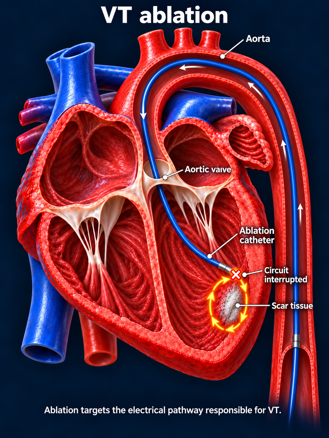
In selected patients with recurrent VF, ablation can target a premature ventricular beat, or PVC, that repeatedly triggers VF. This is a specialized treatment rather than an option for every patient with VF.
Updated October 2026. Educational information; your own care plan may differ. Heart illustrations and EKG examples are simplified for teaching.