What should I do now?
Call 911
Call 911 for collapse, fainting with racing, chest pressure, severe trouble breathing, or repeated ICD shocks. If someone is unresponsive and not breathing normally, follow the dispatcher's CPR/AED instructions.
Contact your care team
After one ICD shock with complete recovery, contact your device team promptly and follow your personal plan. Report recurrent racing or new symptoms.
Keep your care plan
Keep your treatment and follow-up plan. Bring questions, readings, and your medication list.
SECTION 1
What are ventricular tachycardia and ventricular fibrillation?
Ventricular tachycardia (VT) and ventricular fibrillation (VF) are abnormal heart rhythms that start in the heart’s lower chambers, called the ventricles. These chambers pump blood to your lungs and the rest of your body.
Both rhythms disrupt the heart’s normal electrical activity, but they affect the heartbeat differently.
How does the heart normally beat?
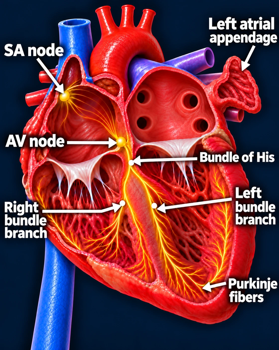
Follow one normal heartbeat
SA node → Atria → AV node → Ventricles
- The SA node starts the signal. The heart’s natural pacemaker in the right upper chamber begins each normal sinus beat.
- The signal spreads through both atria. The upper chambers contract, moving blood into the ventricles.
- The AV node briefly delays the signal. This pause gives the ventricles time to finish filling.
- The signal activates the ventricles. It travels through the bundle of His, right and left bundle branches, and Purkinje fibers. The lower chambers contract, pumping blood to the lungs and body.
The atria contract first; the ventricles follow. This organized sequence produces a coordinated heartbeat.
What is ventricular tachycardia?
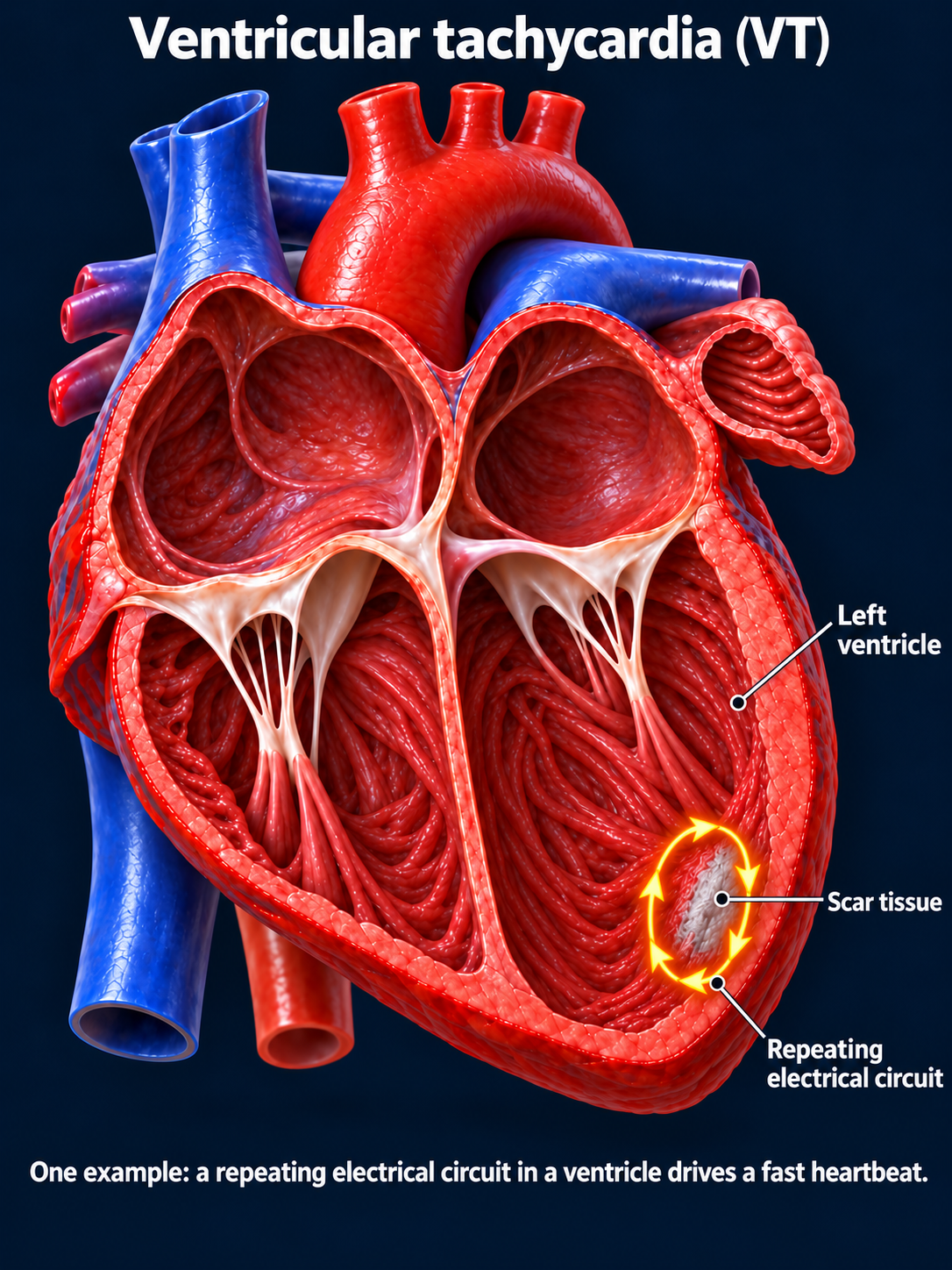
During VT, abnormal electrical signals in the ventricles take over and make the lower chambers beat rapidly. The heart may have less time to fill between beats and may pump less blood to the brain and body.
Some episodes stop on their own after a brief run. Others continue and require treatment to stop them. Doctors call these nonsustained VT and sustained VT.
The seriousness of VT depends on the rhythm, how long it lasts, and the condition of your heart. Some episodes are tolerated, while others cause a dangerous drop in blood pressure, fainting, or cardiac arrest. VT can also deteriorate into VF.
What is ventricular fibrillation?
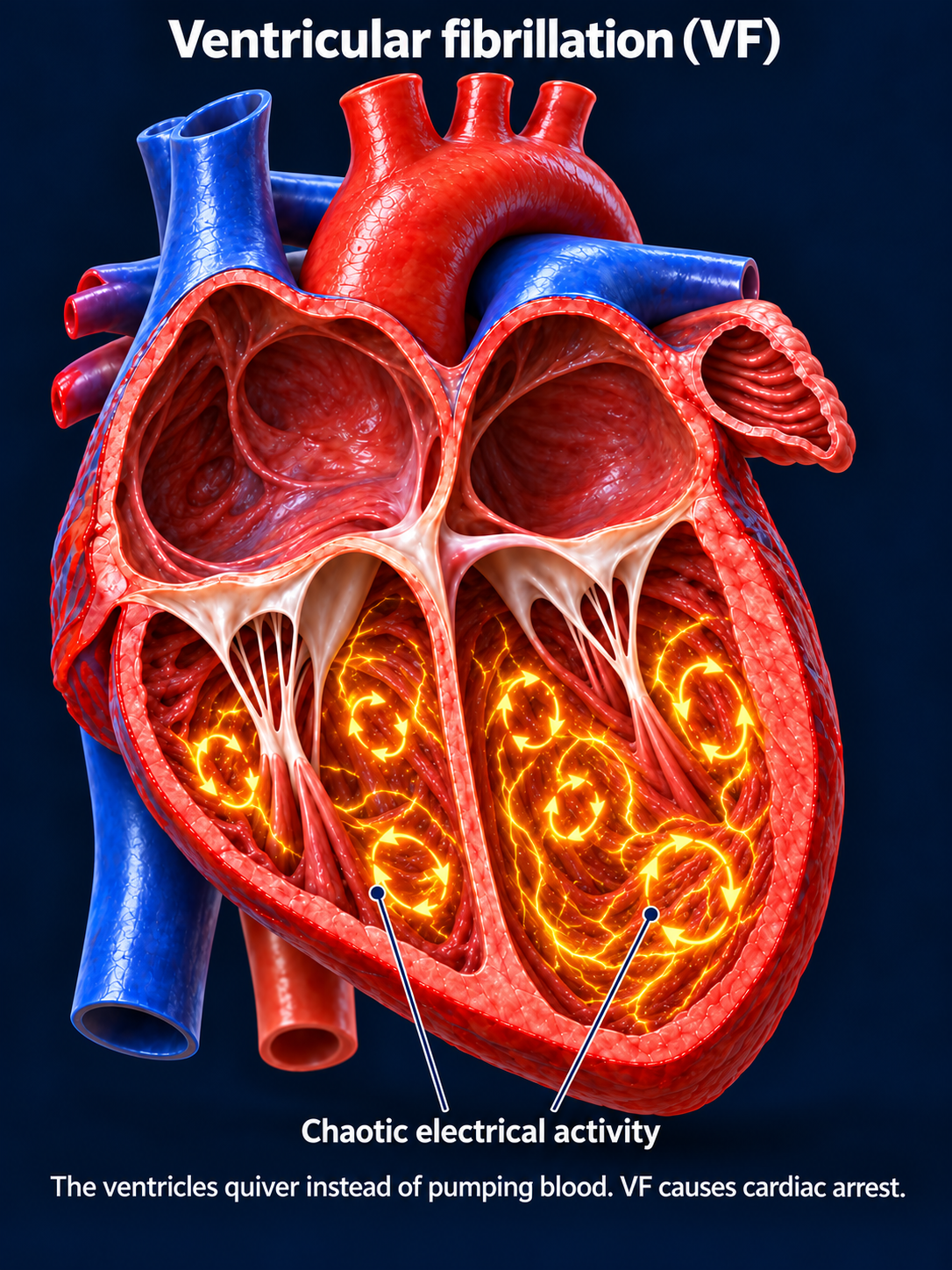
During VF, electrical activity in the ventricles becomes chaotic. Instead of squeezing together, the lower chambers quiver and cannot pump blood effectively.
VF causes cardiac arrest. Blood flow to the brain and body stops, and the person becomes unconscious. Immediate CPR and a shock from a defibrillator are needed to give the person the best chance of survival.
How do these rhythms start?
- Scar tissue, such as damage from a previous heart attack.
- A weakened or diseased heart muscle.
- Reduced blood flow to the heart.
- Certain medications or electrolyte imbalances.
- Inherited conditions that affect the heart’s electrical system.
Some types of VT can also occur in people without obvious heart muscle damage. Finding the cause helps your care team choose the right treatment.
Updated October 2026. Educational information; your own care plan may differ. Heart illustrations and EKG examples are simplified for teaching.