What should I do now?
Call 911
Call 911 for collapse, fainting with racing, chest pressure, severe trouble breathing, or repeated ICD shocks. If someone is unresponsive and not breathing normally, follow the dispatcher's CPR/AED instructions.
Contact your care team
After one ICD shock with complete recovery, contact your device team promptly and follow your personal plan. Report recurrent racing or new symptoms.
Keep your care plan
Keep your treatment and follow-up plan. Bring questions, readings, and your medication list.
SECTION 3
How are VT and VF diagnosed?
VT and VF are diagnosed by recording the heart’s electrical activity. After identifying the rhythm, your care team evaluates what caused it and how well your heart is working.
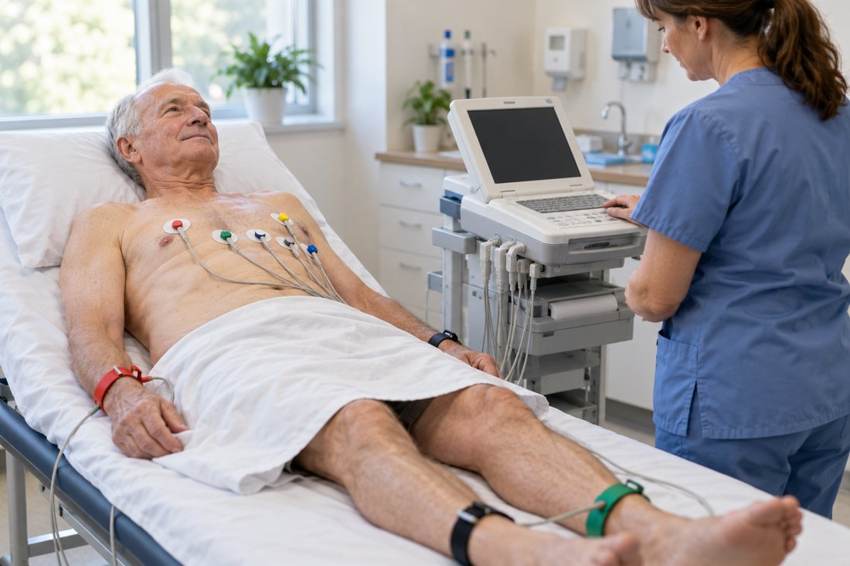
An EKG or rhythm strip
An electrocardiogram, called an EKG or ECG, records electrical signals through small stickers placed on your skin.
An EKG obtained during an episode can identify VT. Hospital heart monitors can also capture episodes as they happen.
VF is usually identified on an emergency monitor or defibrillator during cardiac arrest. Emergency treatment begins immediately; it should not be delayed for additional testing.
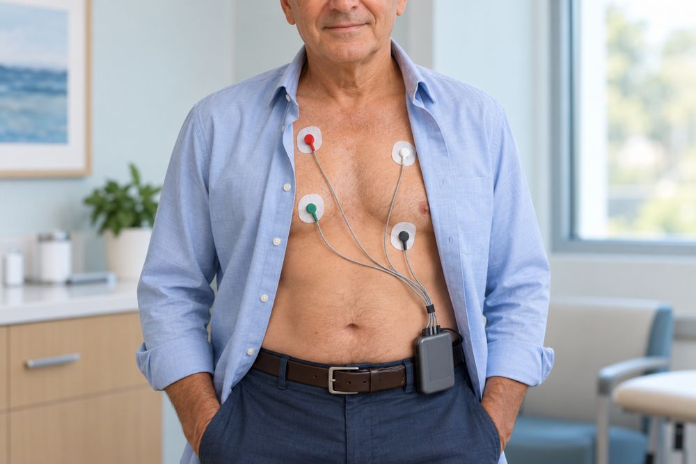
A heart monitor for episodes that come and go
A short office EKG may miss an intermittent rhythm. Your doctor may recommend a Holter monitor, patch monitor, or event monitor to record your heartbeat over a longer period.
Keeping a diary of symptoms and activities helps your care team match what you felt with the recorded rhythm. Selected patients may need a longer-term implanted monitor.
Recordings from an implanted device
If you already have an implantable cardioverter-defibrillator (ICD), your device team can review stored electrical recordings to understand an episode and any treatment the device delivered.
Finding the underlying cause
Your doctor may recommend:
- An echocardiogram: An ultrasound that evaluates the heart’s structure and pumping function.
- Blood tests: To check electrolytes and other possible contributing problems.
- A cardiac MRI: To look more closely at heart muscle damage or scar tissue.
- Tests of the heart’s blood supply: Such as a stress test or coronary angiogram when reduced blood flow or an artery blockage is suspected.
- An electrophysiology study: In selected patients, an EP specialist uses catheters to study the heart’s electrical activity.
- Genetic testing: When an inherited heart condition is suspected.
Not every patient needs every test. Your symptoms, rhythm recordings, medical history, and family history guide the evaluation.
Can a watch or home monitor diagnose VT or VF?
A fast pulse reading alone cannot identify which abnormal rhythm is present. Some watches can record an electrical tracing that may help your doctor, but the recording needs professional review.
Do not rely on a watch, blood pressure cuff, or finger oxygen monitor to rule out a dangerous rhythm or decide whether to seek emergency care.
Updated October 2026. Educational information; your own care plan may differ. Heart illustrations and EKG examples are simplified for teaching.