What should I do now?
Call 911
Call 911 for chest pressure, severe trouble breathing, or fainting. Do not drive yourself.
Contact your care team
Contact your care team promptly for increasing breathlessness, swelling, reduced activity, new palpitations, dizziness, or near-fainting.
Keep your care plan
Keep your treatment and follow-up plan. Bring questions, readings, and your medication list.
SECTION 4
How is heart valve disease treated?
Treatment depends on which valve is affected, how severe the problem is, your symptoms, and how the valve is affecting your heart. Some patients need monitoring, while others benefit from valve repair or replacement.
1. Monitoring your valve
Mild valve disease often does not require a procedure. Your cardiologist may recommend regular visits and repeat echocardiograms to watch for changes.
Even if you feel well, keep your follow-up appointments. Significant valve disease can sometimes require treatment before symptoms appear or lasting heart damage develops.
2. Medications to manage symptoms and support the heart
Depending on your condition, medications may help:
- Remove extra fluid: Diuretics, or “water pills,” can reduce swelling and breathlessness.
- Control blood pressure: This can reduce the workload on your heart.
- Treat heart failure or abnormal rhythms: When these occur alongside valve disease.
- Prevent blood clots: Blood thinners are needed for certain conditions, rather than for every valve problem.
Medications generally do not open a calcified, narrowed valve or repair damaged valve leaflets. They help manage the effects of valve disease. Treating heart failure can sometimes reduce leakage caused by enlargement of the heart.
3. Repairing a valve
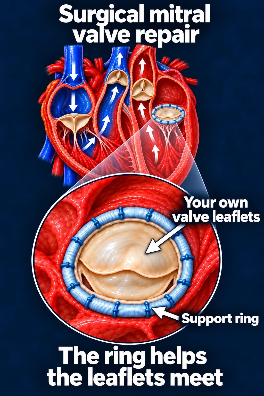
When possible, repair helps your existing valve work better.
A surgeon may reshape valve tissue, repair supporting cords, or place a ring around the valve to help its leaflets meet properly.
Catheter-based repair
For selected patients with significant leakage, a catheter-based repair may be an option. One approach, called transcatheter edge-to-edge repair (TEER), uses a small clip to bring valve leaflets together and reduce leakage. This can be used for selected mitral valve problems; catheter-based treatments are also available for selected tricuspid valve problems.
The best repair depends on the valve’s anatomy and the cause of the leakage.
4. Replacing a valve
If a valve cannot be repaired effectively, it may need replacement.
Surgical valve replacement
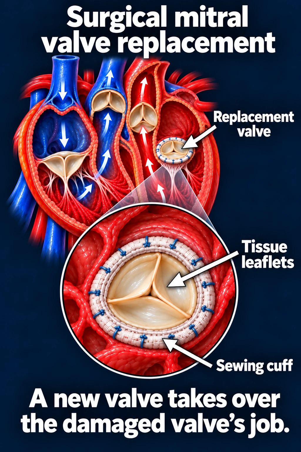
A surgeon removes the diseased valve and replaces it with a new valve. Surgery may be especially helpful when another heart operation is also needed.
Two main replacement types are:
- Mechanical valves: Durable artificial valves that require lifelong warfarin to prevent clots.
- Tissue valves: Made from biological tissue. They can wear out over time and usually do not require lifelong blood thinners solely because of the valve. Another condition, such as AFib, may still require anticoagulation.
Your team helps you choose based on your age, preferences, bleeding risk, and future treatment needs.
Transcatheter aortic valve replacement (TAVR)
For many patients with severe aortic stenosis, TAVR is an alternative to surgical replacement.
A catheter carries a new tissue valve to the heart, usually through an artery in the groin. The new valve expands inside the diseased aortic valve, allowing blood to move forward through the replacement valve.
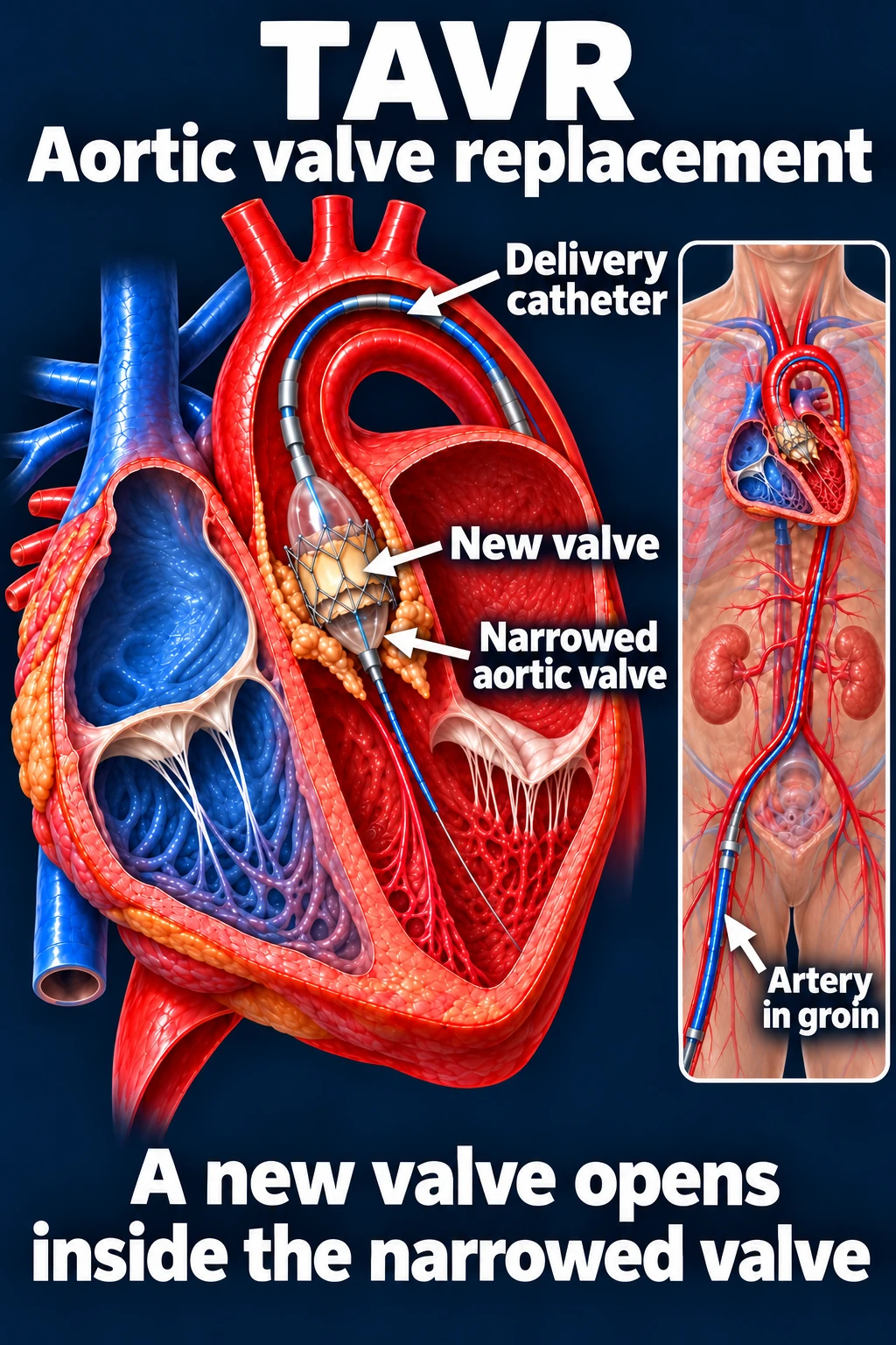
TAVR and surgery each have benefits and risks. Your valve team considers your anatomy, overall health, age, and expected valve durability when recommending an approach.
How do we decide when treatment is needed?
The decision considers more than symptoms alone. Your team reviews the severity of the valve problem, heart size and pumping function, changes over time, and the expected benefit of treatment.
The goal is to treat the valve at the right time—before it causes lasting damage to your heart. A consultation with a specialized valve team can help determine the best procedure and timing.
NIH: Valve disease treatment · AHA: Valve repair · AHA: Valve replacement · NIH: TAVR
Updated October 2026. Educational information; your own care plan may differ. Medical illustrations are simplified examples for teaching.