What should I do now?
Call 911
Call 911 for chest pressure, severe trouble breathing, or fainting. Do not drive yourself.
Contact your care team
Contact your care team promptly for increasing breathlessness, swelling, reduced activity, new palpitations, dizziness, or near-fainting.
Keep your care plan
Keep your treatment and follow-up plan. Bring questions, readings, and your medication list.
SECTION 3
How is heart valve disease diagnosed?
Your doctor reviews your symptoms and medical history, examines you, and uses imaging to assess your valves.
Listening to your heart
A stethoscope may detect a heart murmur—an extra sound caused by blood flow. A murmur may suggest a valve problem, but imaging helps determine its cause and significance.
Echocardiogram: the main test
An echocardiogram, or “echo,” uses ultrasound to create moving pictures of your heart. During a standard echo, a sonographer places a probe on your chest, often while you lie on your left side.
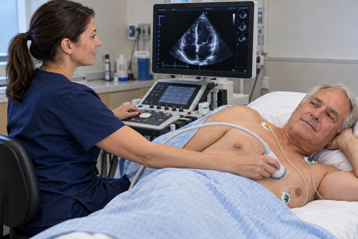
The echo helps your doctor determine:
- Which valve is affected.
- Whether it is narrowed, leaking, or both.
- How severe the problem is.
- How well your heart pumps and whether its chambers have enlarged.
Doppler ultrasound measures blood flow through the valves and helps assess narrowing and leakage.
When are other tests needed?
Not everyone needs additional testing. Depending on the question, your doctor may recommend:
- Transesophageal echo (TEE): An ultrasound probe in the esophagus, behind the heart, provides closer valve views. Your team explains preparation and sedation.
- Cardiac CT: Detailed pictures of the valve, aorta, and surrounding structures, often used to plan a valve procedure.
- Cardiac MRI: Additional assessment of blood flow, leakage, and heart function.
- Supervised exercise testing: In selected patients, to evaluate symptoms during activity.
- Cardiac catheterization: In selected patients, to assess pressures or coronary arteries.
What does “mild, moderate, or severe” mean?
These terms describe the degree of narrowing or leakage. Your doctor combines measurements with symptoms and effects on your heart to guide care.
A normal EKG does not rule out valve disease. An EKG measures electrical activity; an echo evaluates valve structure and blood flow.
Updated October 2026. Educational information; your own care plan may differ. Medical illustrations are simplified examples for teaching.