What should I do now?
Call 911
Call 911 for chest pressure, severe trouble breathing, fainting, or severe dizziness with palpitations. Do not drive yourself.
Contact your care team
Contact your care team for new or increasingly frequent palpitations, reduced activity, or troublesome symptoms.
Keep your care plan
Keep your treatment and follow-up plan. Bring questions, readings, and your medication list.
SECTION 3
How are PACs and PVCs diagnosed?
An EKG or heart monitor records the early electrical beats and helps us determine whether they are PACs or PVCs. We also want to understand how frequently they occur and whether the heart’s structure and pumping function are normal.
An EKG: identifying the early beat
An electrocardiogram, also called an ECG or EKG, records your heart’s electrical activity using small electrodes placed on your chest and limbs.
- A PAC usually shows an early P wave—the electrical signal from the upper chambers—followed by a normal-looking, narrow QRS complex.
- A PVC usually shows an early, wider QRS complex because the electrical signal starts in a ventricle and activates the lower chambers differently.
These are typical patterns. Your clinician reviews the entire tracing to identify the rhythm.
A heart monitor: measuring how often they happen
Premature beats may not occur during a short office EKG. A Holter or patch monitor can record your rhythm during daily activities.
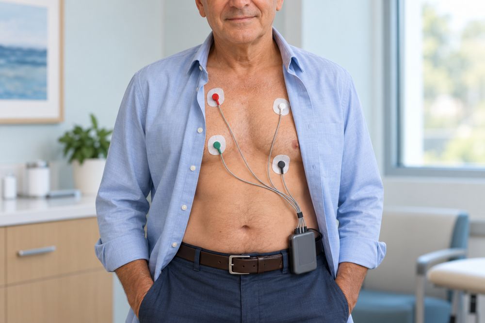
The monitor helps answer:
- Are the premature beats PACs, PVCs, or both?
- How frequently do they occur?
- Do they match the symptoms you report?
- Are there other abnormal rhythms present?
What does “burden” mean?
Burden is the percentage of recorded heartbeats that are premature.
For example, a 10% PVC burden means that approximately 10 out of every 100 recorded heartbeats are PVCs.
A higher PVC burden can increase concern about weakening the heart muscle, but there is no single percentage that determines treatment for every patient. We also consider symptoms, pumping function, and the characteristics of the PVCs.
An echocardiogram: checking the heart’s function
An echocardiogram, or echo, uses ultrasound to examine the heart’s chambers, valves, and pumping strength.
It helps us determine whether an underlying heart condition is present or whether frequent PVCs may be affecting the heart’s function.
Will I need other tests?
Depending on your history and findings, your clinician may recommend blood tests to check electrolytes or thyroid function. Some patients need an exercise stress test or a cardiac MRI to investigate further.
Not everyone needs every test. The evaluation is tailored to your symptoms and rhythm recordings.
Can my smartwatch help?
A watch or handheld device that records an actual EKG may capture an early beat. Save the tracing and share it with your care team.
A heart-rate number alone cannot reliably distinguish PACs from PVCs or measure their burden. An occasional watch recording also cannot replace a continuous monitor when we need to measure how often the beats occur.
The key idea: An EKG identifies the premature beat, a monitor measures its frequency, and an echo helps assess the health of the heart.
Updated October 2026. Educational information; your own care plan may differ. Heart illustrations and EKG examples are simplified for teaching.