Recognize reflex, orthostatic, and cardiac causes of syncope.
What is syncope?
Syncope is a brief loss of consciousness from reduced blood flow to the brain, with spontaneous recovery. Presyncope describes feeling about to faint without losing consciousness. Both deserve assessment in context.
Syncope is a symptom, not a diagnosis. The key question is whether the event reflects a reflex, an orthostatic BP problem, or a cardiac cause.[1]
Reflex / vasovagal syncope
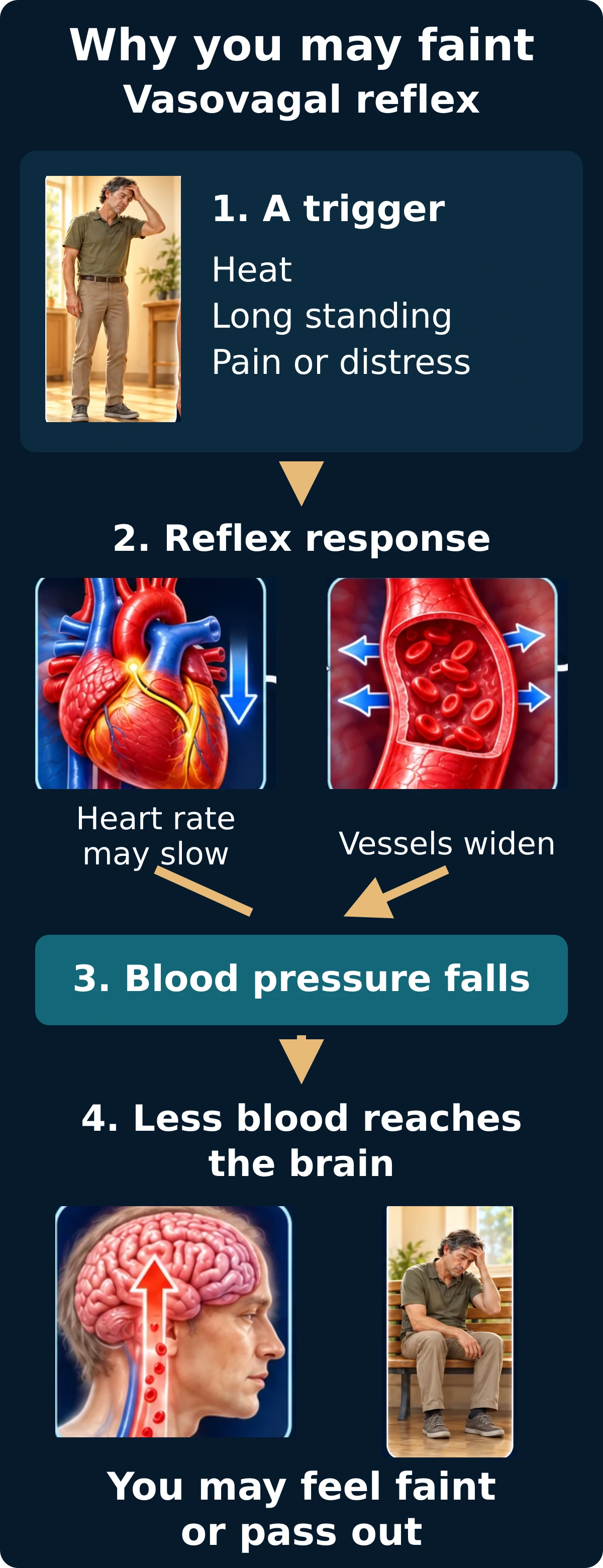
A reflex can lower vascular tone and sometimes slow the heart. Common triggers include prolonged standing, heat, pain, or emotional distress. Warmth, nausea, sweating, or dimming vision may precede the event.
Vasovagal episodes may involve vasodilation, heart-rate slowing, or both. These clues support assessment but do not prove the cause.Open full-size image →
Situational episodes may occur with coughing, urination, or other triggers.[1]
Orthostatic hypotension
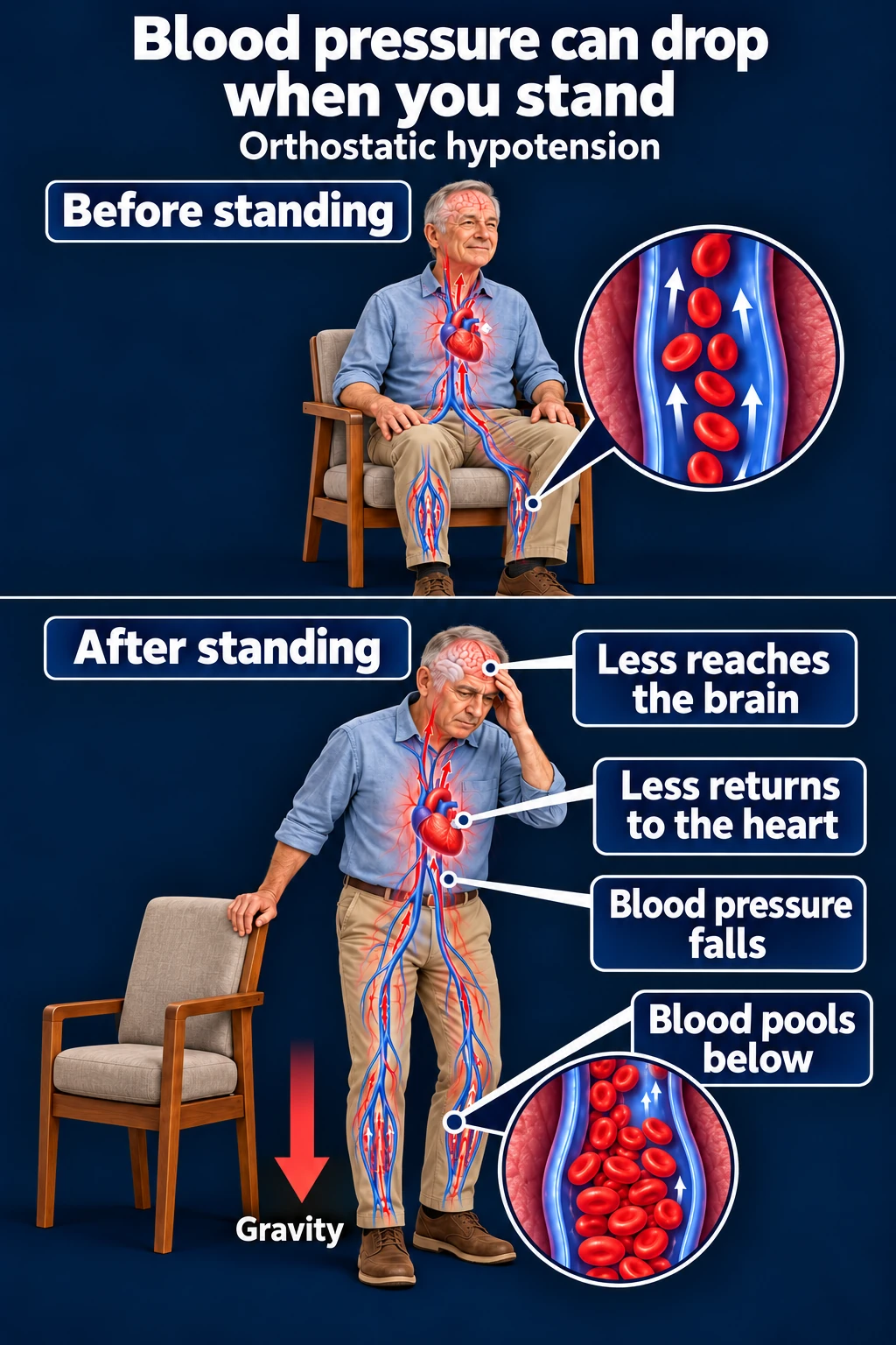
On standing, blood shifts toward the legs and abdomen. If compensation is inadequate, BP falls. Dehydration, medications, or autonomic dysfunction can contribute.
Classical orthostatic hypotension is a sustained fall in systolic BP ≥20 mmHg or diastolic BP ≥10 mmHg within three minutes of standing. The history and symptom response still matter; other orthostatic patterns exist.[3]
A positional BP problem can cause presyncope or syncope. A normal isolated reading does not capture every intermittent or delayed pattern.Open full-size image →
Cardiac syncope
Bradycardia, pauses/AV block, tachyarrhythmias, or structural obstruction can abruptly reduce output. Severe aortic stenosis and cardiomyopathy are examples of structural concerns.
Sinus node dysfunction: The heart’s natural pacemaker starts signals too slowly or pauses.Heart block: Electrical signals from the upper chambers are delayed or blocked on their way to the lower chambers.
Two examples of arrhythmic mechanisms. AV block may arise at the AV node or in the His–Purkinje system; this diagram does not localize every case.
Higher-risk clues
Syncope during exertion, while supine, with sudden palpitations or little warning, abnormal EKG, known structural disease, or a family history of premature sudden death calls for careful cardiac assessment.[4]
Not every blackout is syncope
Clarify whether there was true loss of consciousness and obtain a witness account. Seizure, hypoglycemia, intoxication, and other conditions can mimic syncope. Persistent confusion or focal neurologic findings should broaden the evaluation.[2]