← All clinician topics

CLINICIAN PVC & VT GUIDE · SECTION 1 OF 3

Understanding PVCs & VT

Recognize ventricular ectopy, VT patterns, and the importance of underlying heart disease.

Rhythms from the lower chambers

A premature ventricular contraction (PVC) is an early beat arising in a ventricle. Ventricular tachycardia (VT) is a fast sequence of ventricular beats. These rhythms range from occasional ectopy to life-threatening arrhythmias.

Patients may describe skipped beats, thumps, racing, dizziness, or fainting. Risk depends on the rhythm and the underlying heart: coronary disease, scar, cardiomyopathy, inflammation, and inherited electrical disorders all matter.[1][2]
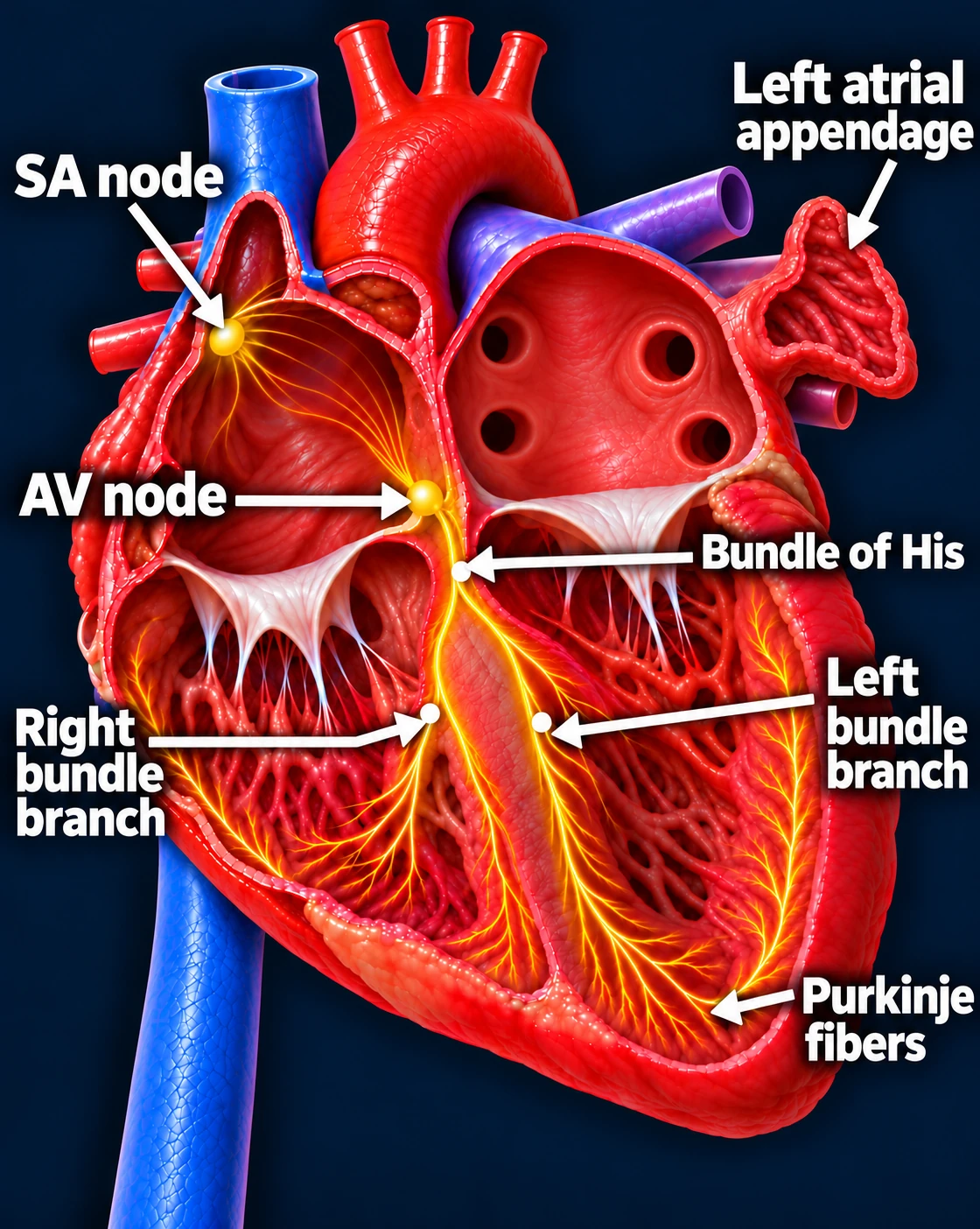
Labeled cutaway heart showing the SA node, AV node, bundle of His, right and left bundle branches, Purkinje fibers, and left atrial appendage.

Follow one normal heartbeat

SA node → Atria → AV node → Ventricles

  1. The SA node starts the signal. The heart’s natural pacemaker in the right upper chamber begins each normal sinus beat.
  2. The signal spreads through both atria. The upper chambers contract, moving blood into the ventricles.
  3. The AV node briefly delays the signal. This pause gives the ventricles time to finish filling.
  4. The signal activates the ventricles. It travels through the bundle of His, right and left bundle branches, and Purkinje fibers. The lower chambers contract, pumping blood to the lungs and body.

The atria contract first; the ventricles follow. This organized sequence produces a coordinated heartbeat.

Normal activation begins in the sinus node and travels to the ventricles.
Open larger image (new tab)
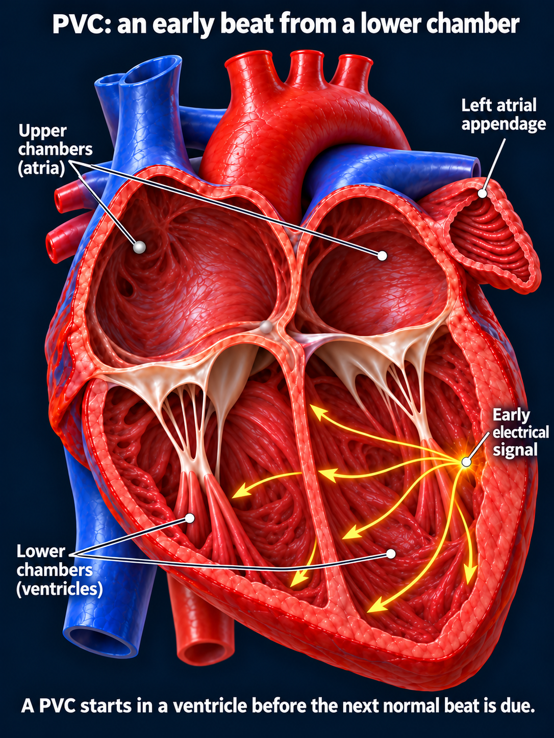
Premature electrical activation originating in a ventricle
A PVC begins in a lower chamber before the next expected sinus beat.
Open full-size image →

Learn the vocabulary

Bigeminy / trigeminy

Every second / third beat is a PVC.

Couplet

Two consecutive ventricular beats.

Nonsustained VT (NSVT)

At least 3 consecutive ventricular beats at >100/min, ending spontaneously in <30 seconds.

Sustained VT

VT lasting >30 seconds, or requiring earlier termination because of hemodynamic compromise.

Monomorphic / polymorphic

Similar / changing QRS morphology during VT.

[3]

Recognize the EKG pattern

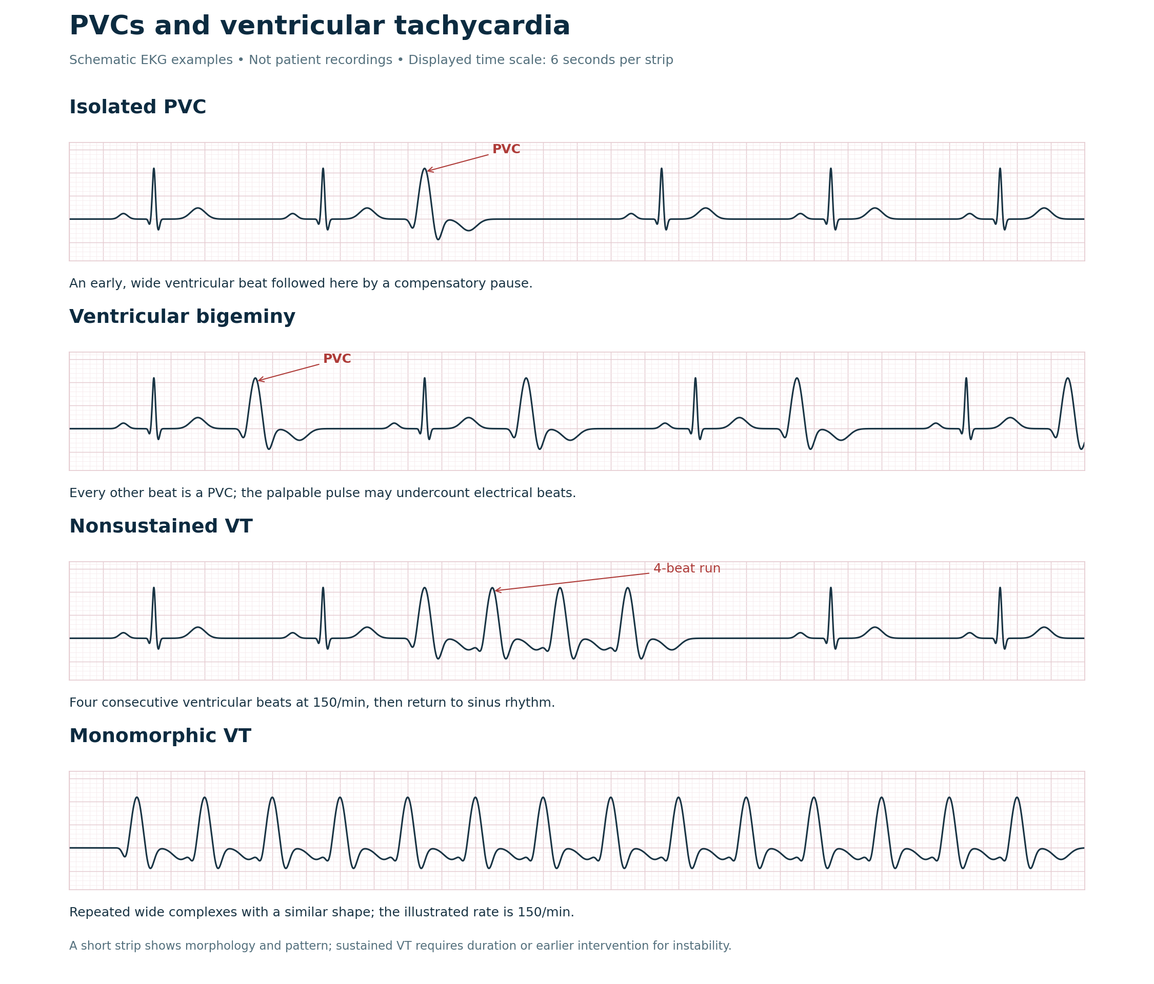
Four schematic EKG strips showing an isolated PVC, ventricular bigeminy, a four-beat run of NSVT, and monomorphic VT
Teaching examples with white backgrounds. The last strip illustrates VT morphology, not a full sustained episode. Confirm findings on the actual tracing; a monitor label alone is insufficient.
Open full-size image →

PVCs usually have a wide QRS without a preceding conducted sinus P wave. During VT, AV dissociation, capture beats, or fusion beats can support the diagnosis, but their absence does not exclude it.

Why PVC burden matters

Burden = PVCs ÷ total beats. Frequent PVCs may weaken ventricular function. An unexplained reduced EF with a burden around ≥10% warrants assessment for PVC-induced cardiomyopathy; this is not a universal cutoff or an automatic ablation indication. PVCs may also be a consequence of existing disease.[4]

Polymorphic VT and torsades

Torsades de pointes is polymorphic VT associated with a prolonged QT interval between episodes. Other polymorphic VT can occur with a normal QT, including during acute ischemia. Both require urgent assessment.[5]