Determine urgency, gather the evidence, and coordinate appropriate testing.
Start with urgency
Ongoing or recurrent concerning chest discomfort, new rest symptoms, marked worsening, syncope, hemodynamic instability, or severe dyspnea warrants emergency assessment. Activate EMS when ACS or another serious acute cause is suspected; do not send the patient to an outpatient stress test.
Obtain an EKG promptly when feasible, without delaying transfer. A normal initial EKG or a prior reassuring test does not exclude a new ACS.[1], [2]
Clarify the history and examine the patient
Document onset, duration, exertional/rest pattern, radiation, associated symptoms, relief, and change from baseline. Record BP, pulse, oxygen saturation, and signs of heart failure or poor perfusion.
Review prior CAD/MI/PCI/CABG, risk factors, current medications, and adherence. Consider aortic dissection, pulmonary embolism, and other urgent diagnoses when the presentation suggests them.[1]
Understand what the acute evaluation answers
Emergency pathways combine symptoms, EKGs, serial high-sensitivity troponin, and structured risk assessment. A single elevated troponin indicates myocardial injury; interpretation of its change and evidence of ischemia determines whether it represents MI. Kidney disease, heart failure, sepsis, and other conditions can also cause injury.
Outpatient bloodwork should not delay emergency evaluation of suspected ACS. A single reassuring result outside a validated pathway should not be used to dismiss concerning symptoms.[2]
For stable symptoms, choose testing to answer the question
CCTA
Is coronary plaque/narrowing present, and what is the anatomy?
Stress imaging: echo, nuclear perfusion/PET, or CMR
Is there inducible ischemia, and how extensive is it?
Exercise EKG in selected patients
Are symptoms/EKG changes provoked by exercise when the resting tracing is interpretable?
Cardiology selects the approach using clinical risk, known CAD, prior tests, exercise capacity, resting EKG, renal function, and local expertise. Low-risk stable patients may not need testing; high-risk findings may require angiography.[1]
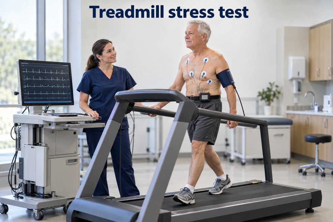
Illustrative stable-patient evaluation. Exercise testing is one option; suspected ACS requires an acute care pathway.Open full-size image →
Make the referral actionable
Send the symptom timeline, EKG, medication list, prior angiography/PCI/CABG details, and available imaging results. Specify whether symptoms are new, worsening, or persistent despite treatment.
Persistent symptoms with nonobstructive coronary arteries may need evaluation for microvascular dysfunction or vasospasm. An anatomical test does not answer every ischemia question.[1]
Clinical takeaway: Determine urgency first. In stable patients, coordinate a risk-based evaluation that answers a clear clinical question.