Understand plaque, ischemia, and the symptom pattern.
What is coronary artery disease?
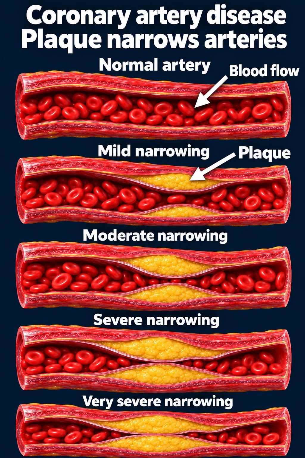
The coronary arteries supply oxygen-rich blood to the heart muscle. In coronary artery disease (CAD), atherosclerotic plaque develops within their walls. Plaque may narrow the channel for blood flow or become disrupted, allowing a clot to form.
Ischemia occurs when oxygen supply is insufficient for the heart muscle’s needs. It can produce angina, impaired function, or an acute coronary event.[1]
Plaque burden and narrowing can vary. These are illustrative examples, not stages every patient must pass through.Open full-size image →
CAD is more than a single blockage.
Risk reduction remains important even when symptoms are controlled or one narrowing has been treated.
What does angina feel like?
Patients may describe pressure, tightness, heaviness, squeezing, or burning rather than “pain.” Discomfort may involve the chest, arm, shoulder, jaw, back, or upper abdomen. Exertional breathlessness or unusual fatigue may be an ischemic equivalent.
Ask about the trigger, duration, frequency, associated symptoms, relief, and change from baseline. Chest discomfort is common in both men and women; accompanying symptoms may vary. Use “cardiac,” “possibly cardiac,” or “noncardiac” rather than dismissing symptoms as “atypical.”[2]
Stable angina versus a possible acute coronary syndrome
Stable angina
Typical features: Predictable symptoms with a similar level of exertion or stress, improving with rest or prescribed therapy.
Clinical response: Assess risk and arrange appropriate cardiology evaluation and treatment.
Possible ACS
Typical features: New severe symptoms, symptoms at rest, prolonged discomfort, or a worsening pattern—especially with dyspnea, sweating, nausea, or instability.
Clinical response: Urgent emergency assessment; activate EMS when ACS is suspected in clinic.
The symptom pattern guides urgency; it does not establish the diagnosis. A patient with previously stable angina can develop a new acute event.
How does plaque cause a heart attack?
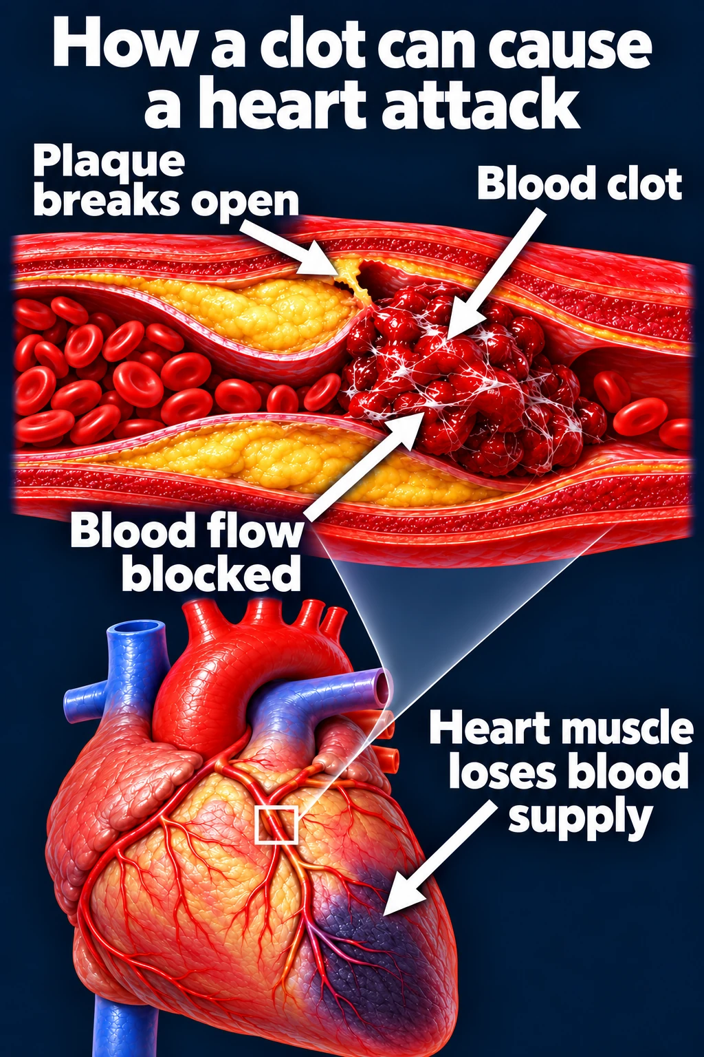
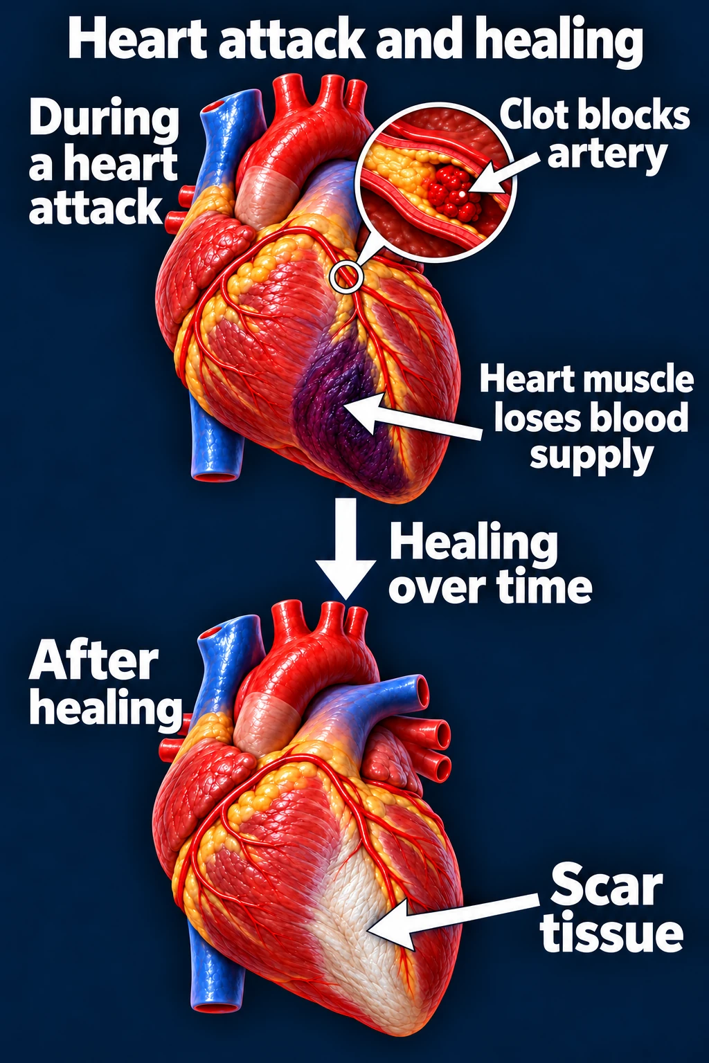
Plaque rupture or erosion can trigger thrombus formation and abruptly reduce coronary blood flow. Prolonged ischemia can injure or kill heart muscle. Acute coronary syndromes include unstable angina, NSTEMI, and STEMI; assessment uses the clinical presentation, EKG, and cardiac biomarkers.[3], [4]
One common mechanism of an acute coronary event: disrupted plaque and thrombosis. Not all myocardial infarctions have this mechanism.Open full-size image →Some infarcted myocardium heals with scar. The extent of injury varies and can affect ventricular function and arrhythmic risk.Open full-size image →
A normal initial EKG does not exclude ACS.
Do not delay emergency evaluation when the history is concerning. Serial EKGs and troponin assessment may be needed in the acute setting.[2]
Angina can occur without a major obstructive lesion
Coronary microvascular dysfunction and vasospasm can cause ischemic symptoms even when the larger arteries do not show a severe fixed narrowing. Persistent symptoms deserve appropriate assessment rather than reassurance based only on the absence of obstructive CAD.[5]
Risk factors and referral
Review LDL cholesterol, smoking, BP, diabetes, family history, and other vascular disease. Known CAD, prior MI, PCI/CABG, and kidney disease influence clinical risk.[1], [5]
Refer suspected CAD, recurrent exertional symptoms, uncertain ischemic symptoms, or angina despite treatment to cardiology. New or worsening symptoms may require acute evaluation rather than routine referral.