Recognize major myocardial patterns and AL versus ATTR amyloidosis.
Cardiomyopathy describes a myocardial disorder
The heart muscle may be enlarged, thickened, scarred, or unusually stiff. Phenotype is a starting point; the underlying cause determines treatment and family implications.
Dilated
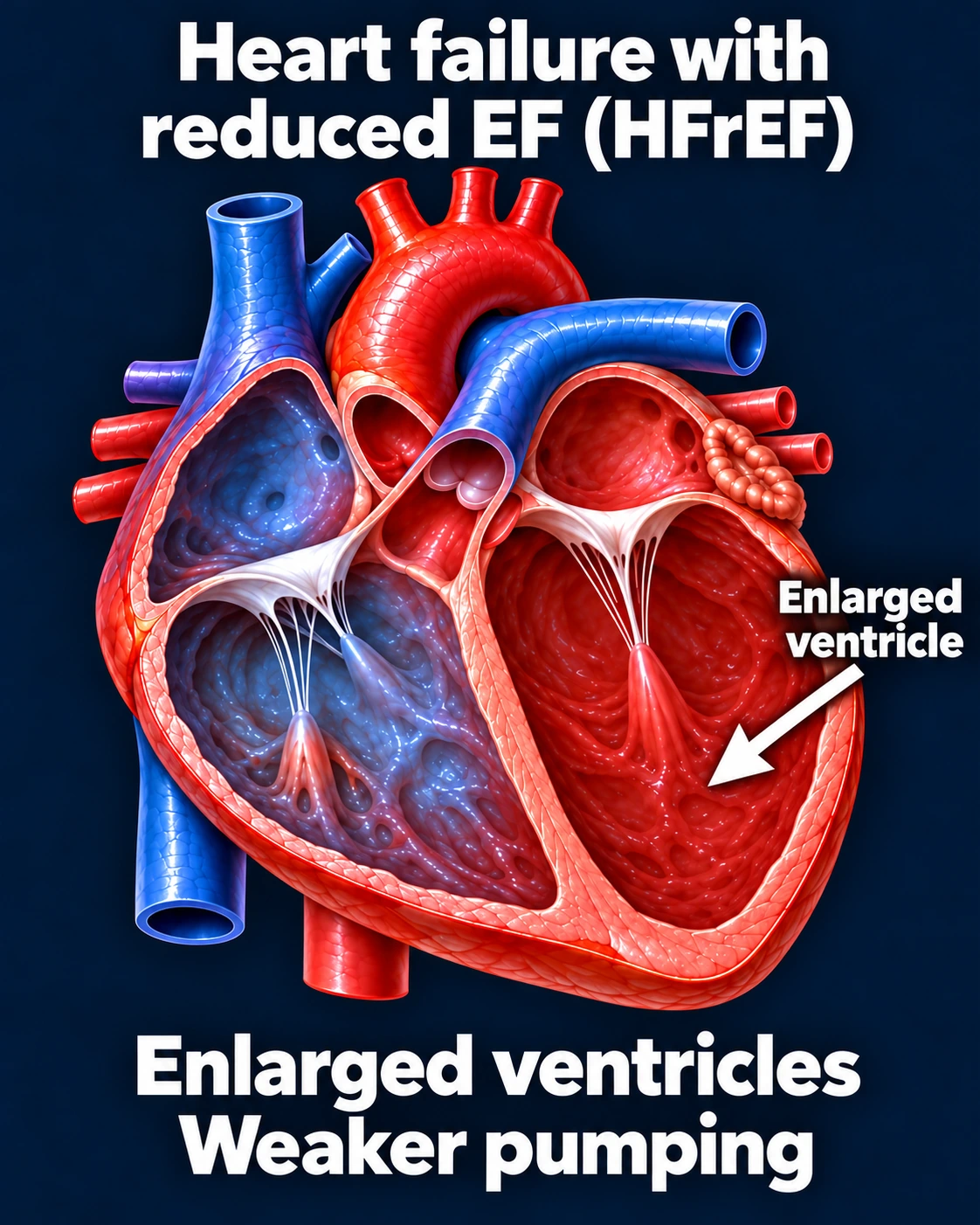
Enlargement and reduced systolic function may occur
Hypertrophic
Unexplained thickening; obstruction and arrhythmic risk vary
Restrictive/infiltrative
Impaired filling; EF may initially remain preserved
Arrhythmogenic
Myocardial disease with important arrhythmic risk
A reduced-EF phenotype example. Appearance alone does not establish the cause.Open full-size image →[1]
Amyloidosis: protein deposition in the heart
AL arises from immunoglobulin light chains associated with a plasma-cell disorder. ATTR arises from transthyretin and can be hereditary or wild type. Distinguishing them promptly is essential because treatments differ.
Deposits can impair relaxation and eventually pumping function, with AF and conduction disease. Preserved EF does not establish benign disease.