SECTION 1
Understanding your device
An implanted heart device monitors your heartbeat and provides treatment when needed. Different devices serve different purposes. Your doctor will explain which device is best for you based on your heart rhythm, pumping strength, and other health factors.
Pacemaker: supporting a slow heartbeat
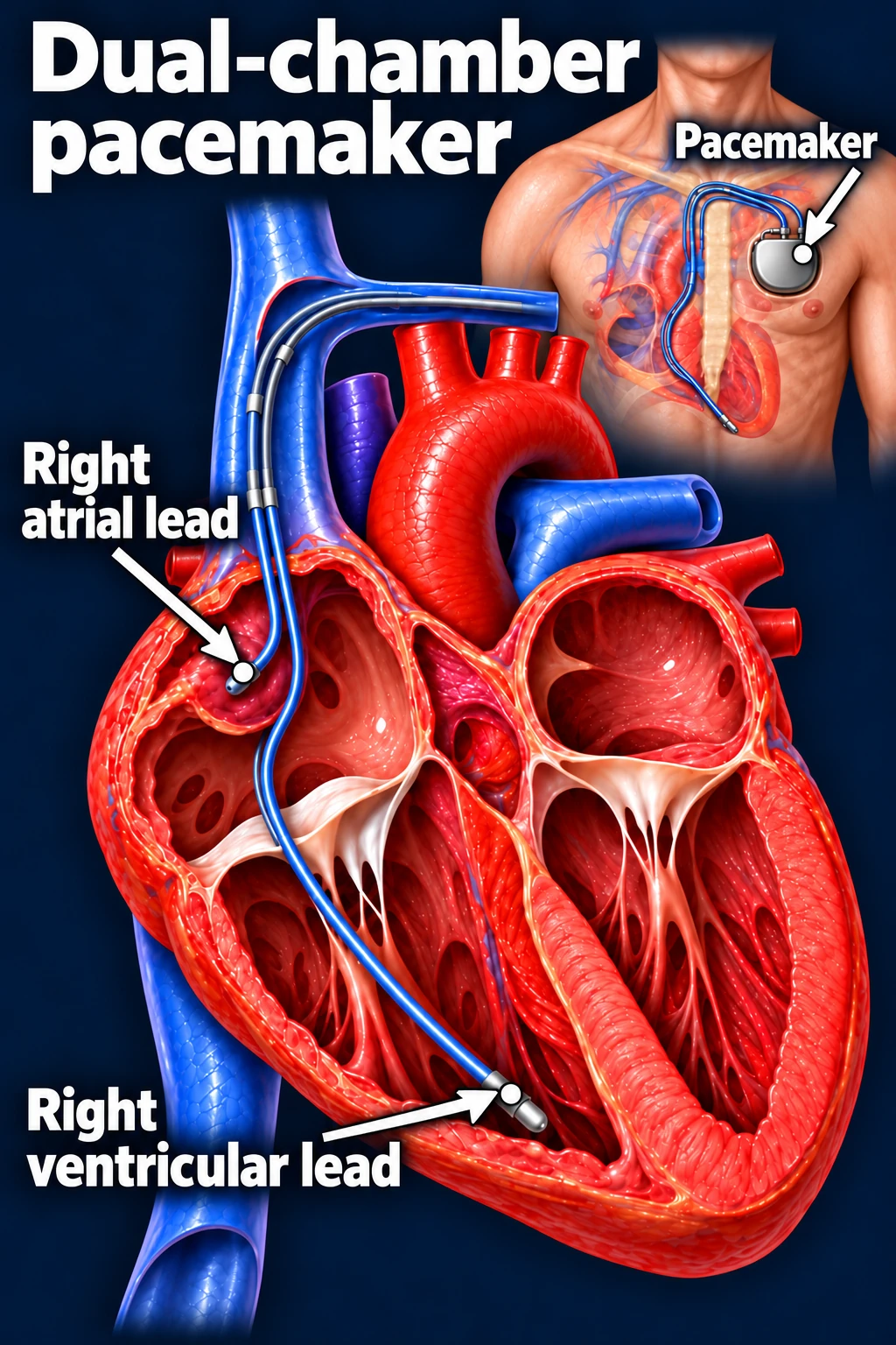
A pacemaker sends small electrical signals when your heartbeat is too slow or signals do not travel properly through the heart. These signals help the heart maintain an appropriate rate.
Most traditional pacemakers have a small battery unit placed under the skin of the upper chest, usually on the left side. Thin insulated wires, called leads, connect the device to the heart.
Defibrillator, or ICD: treating dangerous fast rhythms
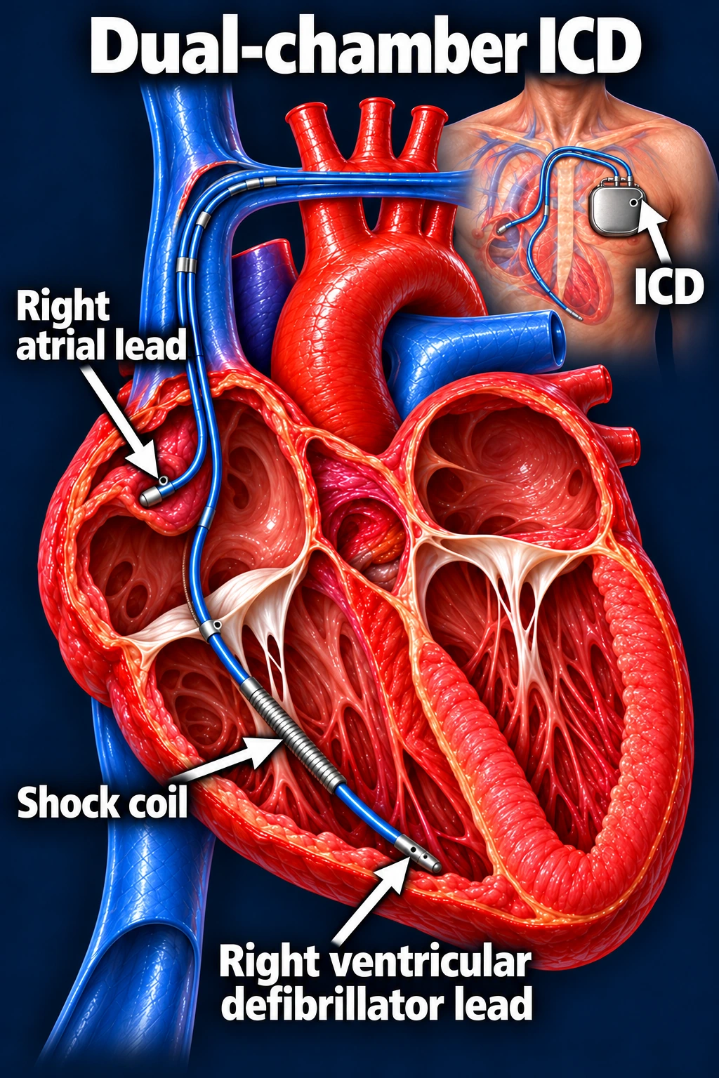
An implantable cardioverter-defibrillator monitors for dangerous rhythms such as ventricular tachycardia and ventricular fibrillation.
Depending on the rhythm and device, an ICD can deliver rapid pacing or an electrical shock to stop the abnormal rhythm. Many ICDs also provide pacing for a slow heartbeat.
CRT: helping the ventricles work together
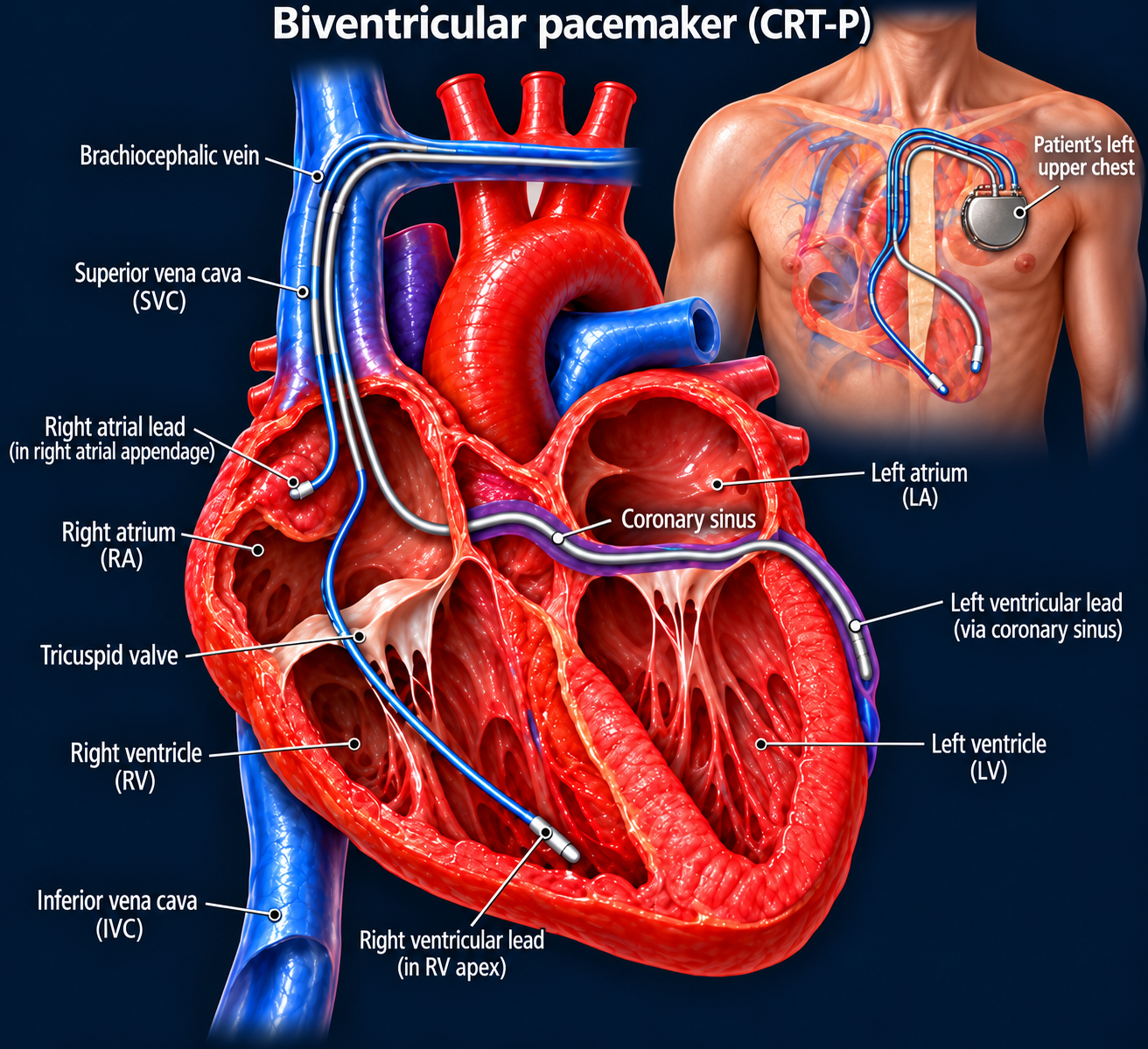
Some patients with heart failure have electrical delays that cause the lower chambers to squeeze out of sync. Cardiac resynchronization therapy, or CRT, helps coordinate their contractions and can improve symptoms and heart function in selected patients.
CRT-P: Provides pacing and resynchronization.
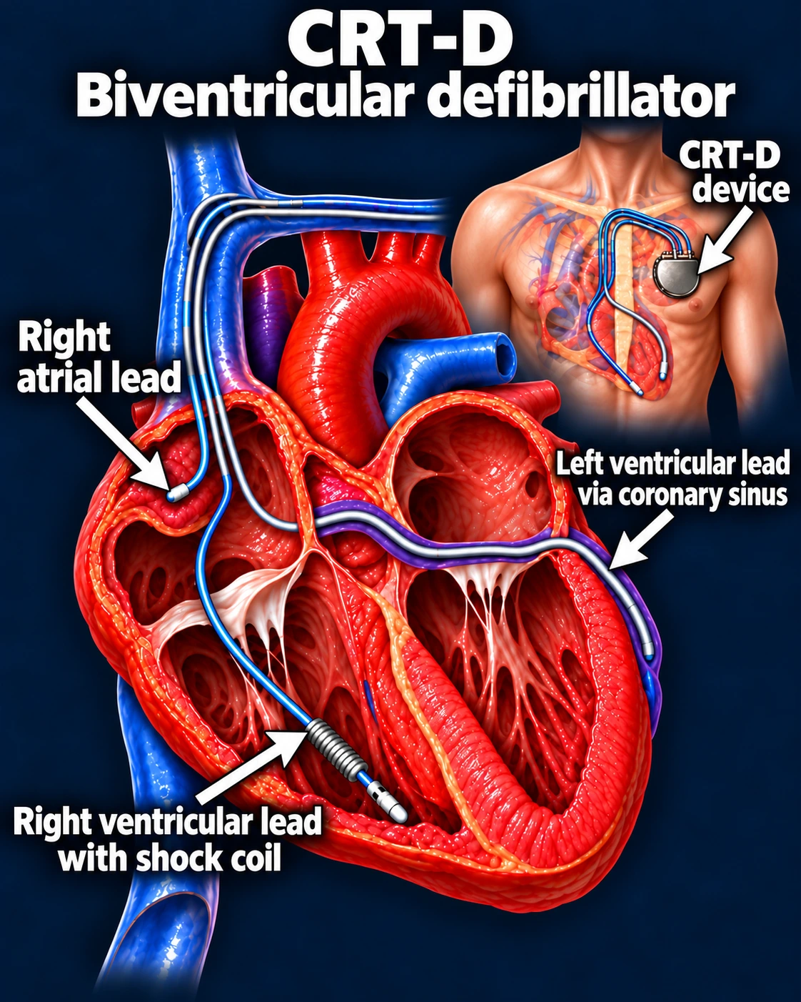
CRT-D: Provides resynchronization plus defibrillator protection.
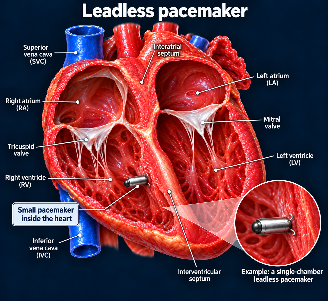
Leadless pacemaker: a device inside the heart
A leadless pacemaker is placed directly inside the heart through a catheter inserted into a vein in the groin. It does not require a chest pocket or traditional leads running from the chest to the heart.
Leadless pacing is suitable for selected patients. Your doctor will explain whether it meets your needs.
Your device is part of your treatment plan
A device can support your heart rhythm or protect against dangerous rhythms, but you may still need medicines and ongoing cardiology care. Its settings are tailored to you and checked regularly after implantation.
Learn about implantable loop recorders for long-term heart rhythm monitoring.
Updated October 2026. Educational information; your own care plan may differ. Medical illustrations are simplified examples for teaching.