Understand valve anatomy, stenosis, regurgitation, and their effects on the heart.
What do the heart valves do?
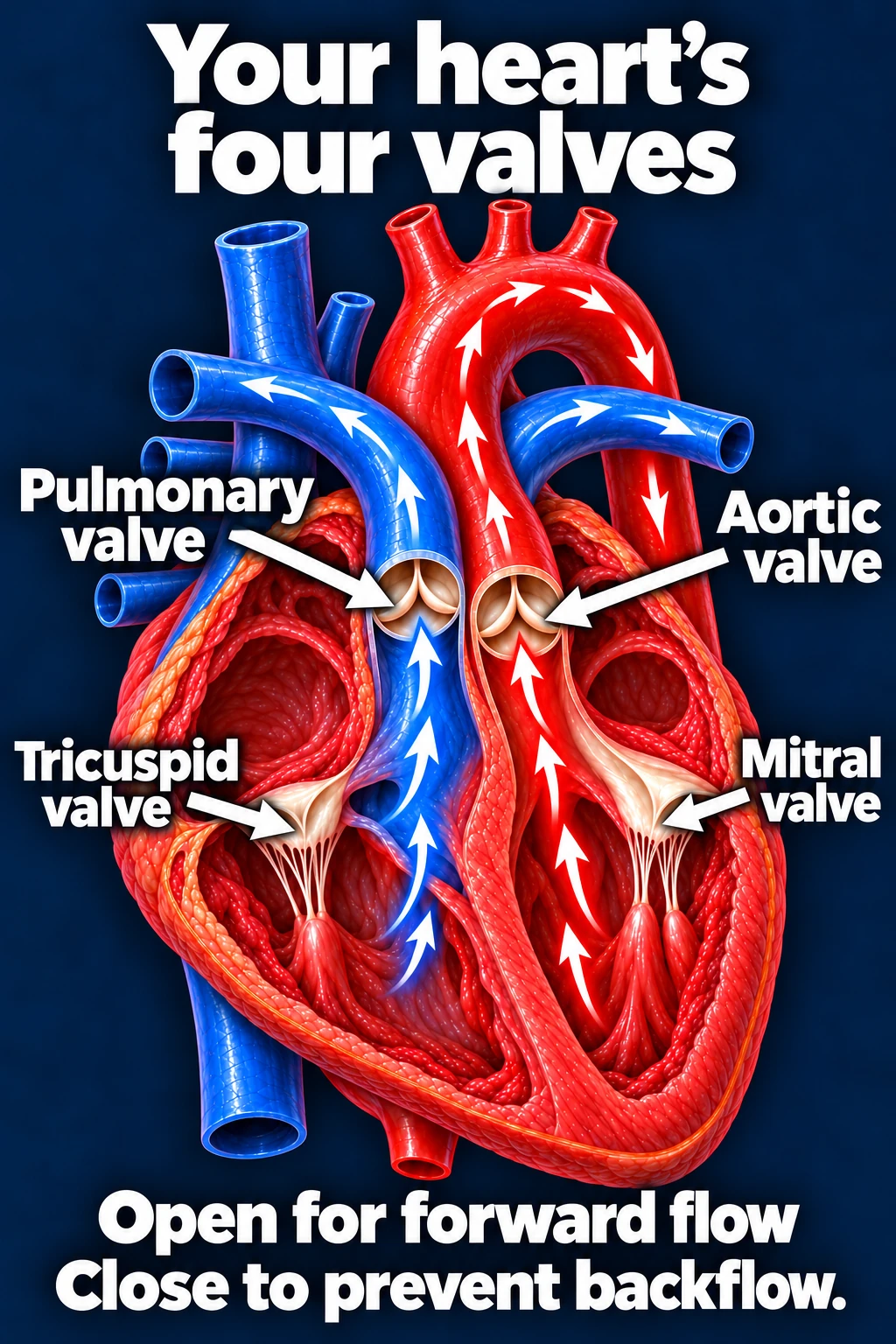
The heart’s four valves keep blood moving in the correct direction. They open to permit forward flow and close to prevent backflow. Disease can impair opening, closure, or both.
Tricuspid
Right atrium → right ventricle
Pulmonary
Right ventricle → pulmonary artery
Mitral
Left atrium → left ventricle
Aortic
Left ventricle → aorta
Simplified valve anatomy and circulation. The valves support forward flow through the right and left heart.Open full-size image →
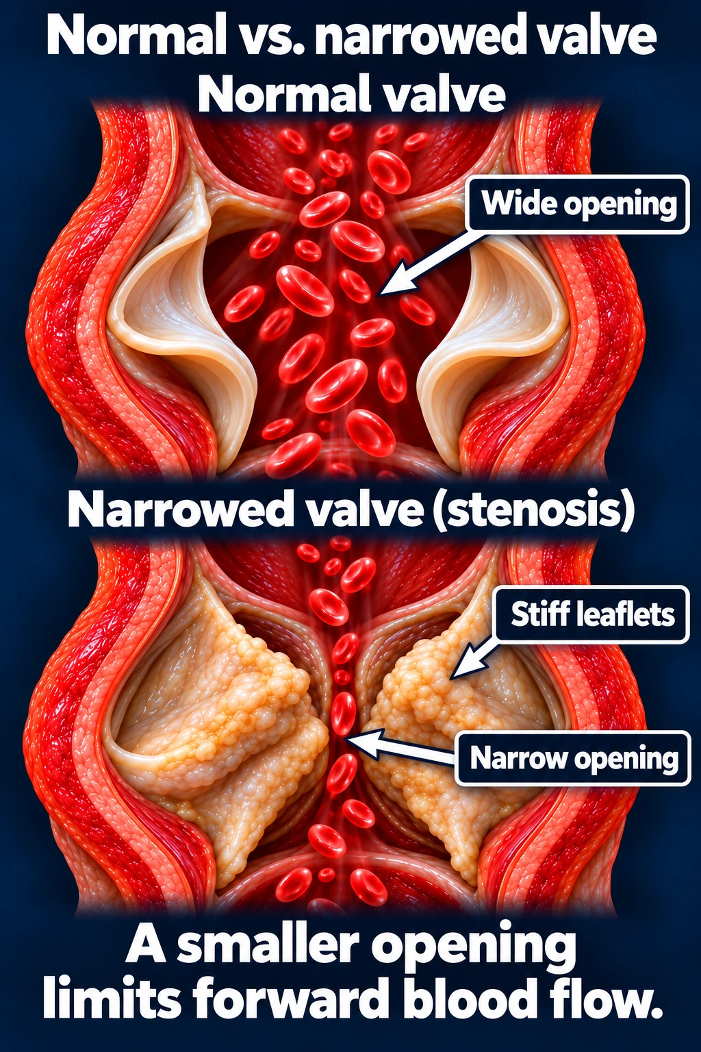
Stenosis · The valve does not open adequately
A narrowed valve obstructs forward flow. The chamber upstream may need higher pressure to move blood through the smaller opening.
Aortic stenosis increases LV workload; mitral stenosis can raise left atrial and pulmonary pressures. Symptoms depend on the affected valve, severity, and the heart’s response.[1]
Illustrative stenosis: restricted opening creates an obstacle to forward flow. Appearance varies by valve and cause.Open full-size image →
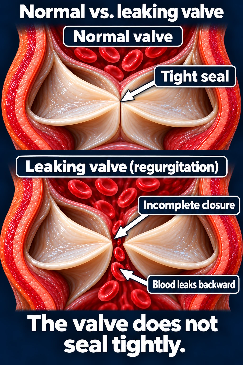
Regurgitation · The valve does not seal adequately
A leaking valve allows blood to flow backward when it should be closed. This adds volume and can raise pressures or enlarge affected chambers over time.
Regurgitation may arise from a diseased valve or from changes in the surrounding chamber/valve apparatus. Acute severe regurgitation can cause rapid deterioration before chamber enlargement has developed.[1], [4]
Illustrative regurgitation. The mechanism, severity, and effect on the heart determine management.Open full-size image →
A murmur is a clue
A murmur is a sound created by blood flow heard during auscultation. Some are innocent; others suggest valve disease or another structural problem. Timing, location, radiation, and accompanying findings help guide assessment.
Auscultation alone does not fully define the lesion or its severity. Echocardiography assesses valve anatomy/function, chamber response, and associated findings; cardiology integrates those results with the history and examination.[2], [4]
Recognize symptoms—even when the patient has adapted
Exertional dyspnea or declining exercise capacity
Fatigue, chest discomfort, or palpitations
Lightheadedness, near-syncope, or syncope
Orthopnea or swelling of the legs/abdomen
Ask what the patient can do now compared with months ago. Walking less, avoiding stairs, or sleeping upright may conceal a gradual decline. Severe valve disease can be present with few reported symptoms.[3]
Severity is more than a symptom count
The affected valve, stenosis versus regurgitation, measured severity, ventricular function/size, pulmonary pressures, and change over time all matter. Some asymptomatic patients still need intervention assessment.
Significant valve disease requires a clear surveillance plan. Severe disease being considered for intervention should be assessed by a multidisciplinary valve team.[4]
Referral principle: Refer a concerning new murmur, symptomatic valve disease, or significant disease without a clear follow-up plan to cardiology. Refer severe disease for timely valve-team assessment.
Urgent symptoms need acute evaluation.
Severe breathlessness, pulmonary edema, syncope with concerning findings, ongoing chest pain, or hemodynamic instability may require emergency care rather than routine referral.