Understand the common mechanisms, recognize the EKG clues, and explain why ablation can be curative.
What is supraventricular tachycardia?
SVT is a fast heart rhythm arising above the ventricles—from the upper chambers (atria) or circuits involving the AV node. An abnormal electrical loop or extra atrial source repeatedly activates the heart, producing a rapid heartbeat. Many episodes begin and end suddenly.[7], [1]
SVT is an umbrella term, not one diagnosis. The three common types covered here are AVNRT (a loop near the AV node), AVRT (a loop using an accessory pathway), and focal atrial tachycardia (an extra source in an atrium). Identifying the type helps explain the EKG and choose the right treatment.[1]
Stroke risk: These common SVTs do not carry the same established clot-related stroke risk as AFib or atrial flutter. Blood thinners are not routinely needed solely for AVNRT, AVRT, or focal atrial tachycardia. Coexisting AFib/flutter or another indication requires its own assessment.[3]
Ablation can provide a complete cure. By eliminating the responsible pathway or focus, catheter ablation can permanently stop the SVT—rather than simply suppressing episodes with medication. Most common re-entrant SVTs are highly amenable to cure; success and recurrence vary by mechanism and location, so cure is not guaranteed for every patient.[8], [2]
AVNRT · A loop near the AV node
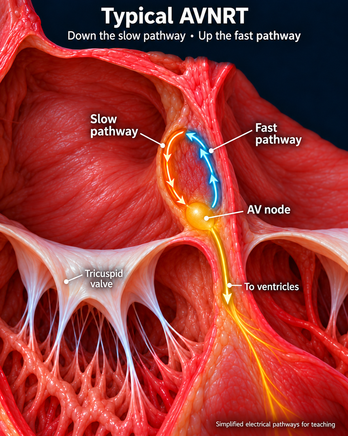
Atrioventricular nodal re-entrant tachycardia uses pathways with different conduction properties in and around the AV node. Typical AVNRT travels down the slow pathway and returns through the fast pathway. Episodes often start and stop abruptly; atrial activation is commonly hidden within the QRS.[1]
Typical AVNRT travels down the slow pathway (orange) and back up the fast pathway (blue). These functional pathways are enlarged and simplified for teaching.Open larger image (new tab)
AVRT · A loop using an accessory pathway
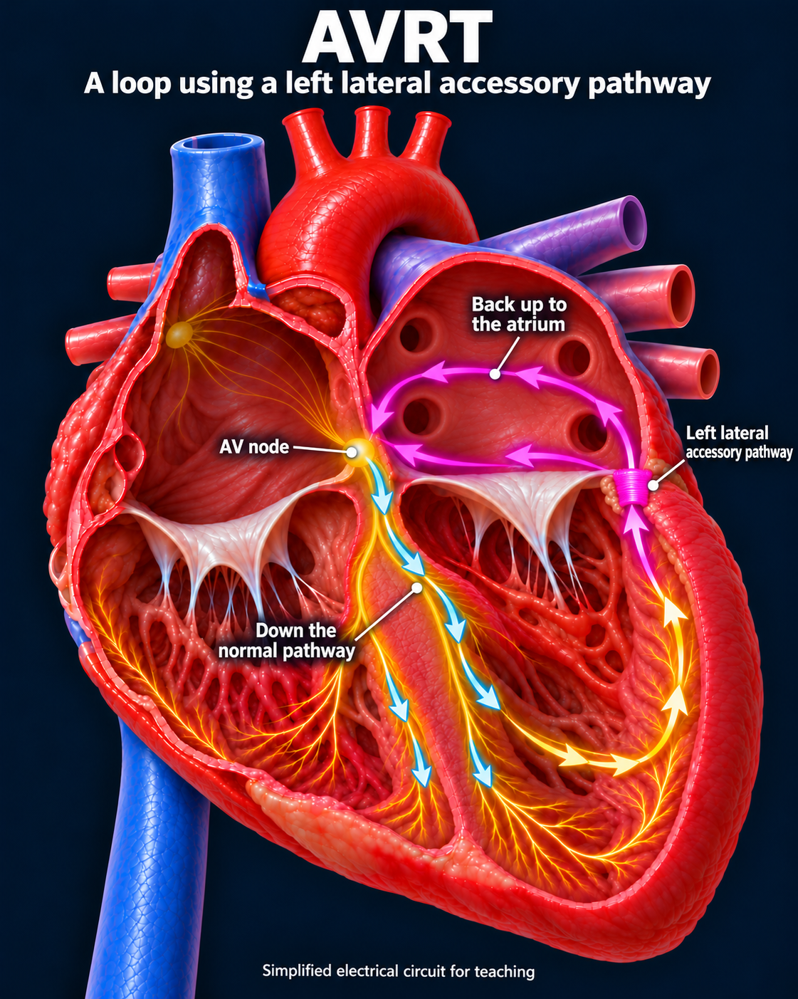
In orthodromic atrioventricular re-entrant tachycardia, the impulse travels down the normal AV conduction system and returns through an accessory pathway. The QRS is usually narrow. A concealed pathway conducts only backward, so the resting EKG may show no delta wave.[2]
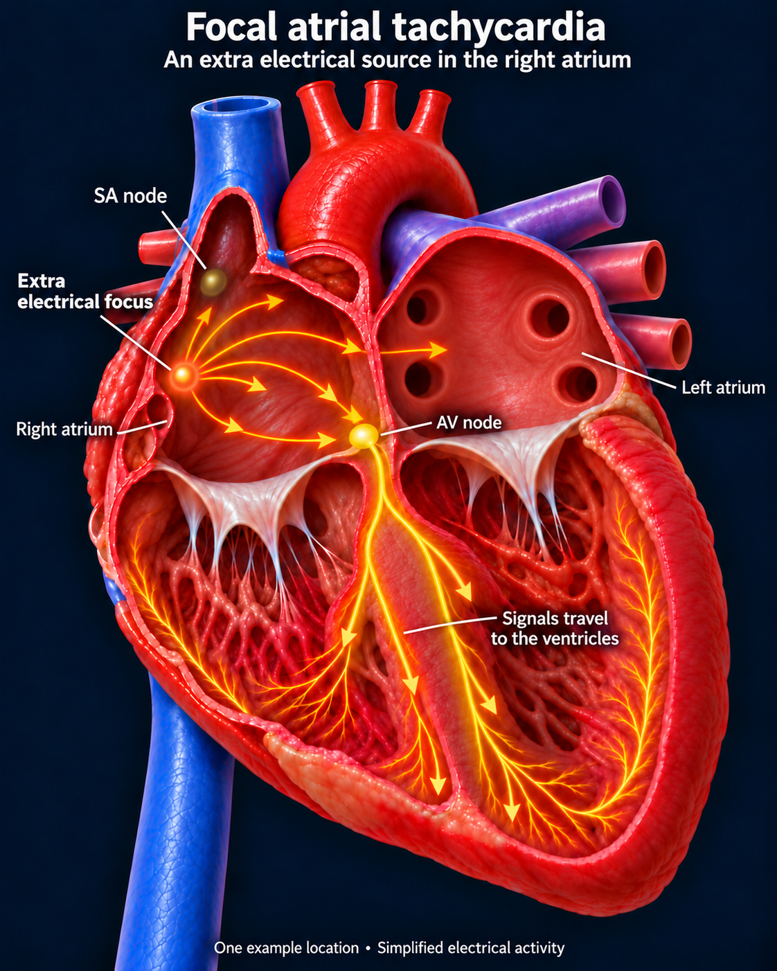
An atrial focus drives the rhythm rather than an AV node–dependent loop. P waves may differ from sinus rhythm. Some episodes gradually accelerate or slow. Persistent tachycardia can weaken ventricular function.[3]
An example right atrial focus; focal atrial tachycardia can arise elsewhere in either atrium.Open larger image (new tab)
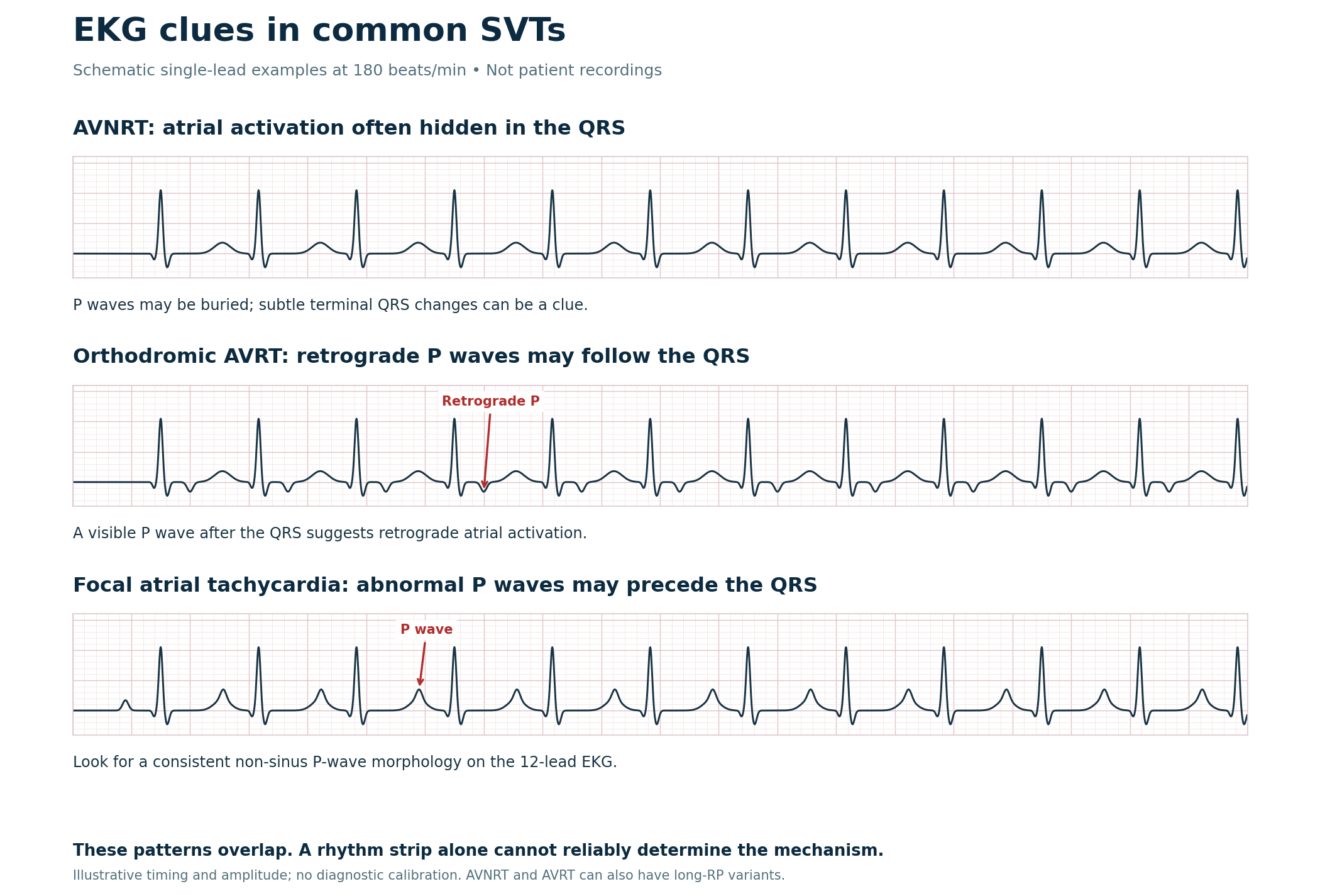
Recognizing SVT on the EKG
Teaching examples only. Interpret P-wave timing and morphology using a 12-lead EKG; these clues do not establish the mechanism. [1, 3]Open full-size image →
Do not diagnose by rate alone. Sinus tachycardia and atrial flutter with 2:1 conduction can mimic SVT. A regular rhythm near 150/min should prompt a search for flutter waves.[3]