Recognize the syndrome, understand EF, and look beyond the number.
What is heart failure?
Heart failure is a clinical syndrome caused by abnormal cardiac structure or function. The heart may have difficulty pumping, relaxing, or filling, leading to elevated pressures, congestion, and sometimes inadequate blood flow to meet the body’s needs.
It can occur with reduced or preserved ejection fraction. Symptoms, signs, and objective cardiac evidence establish the diagnosis; the EF alone does not.[1], [2]
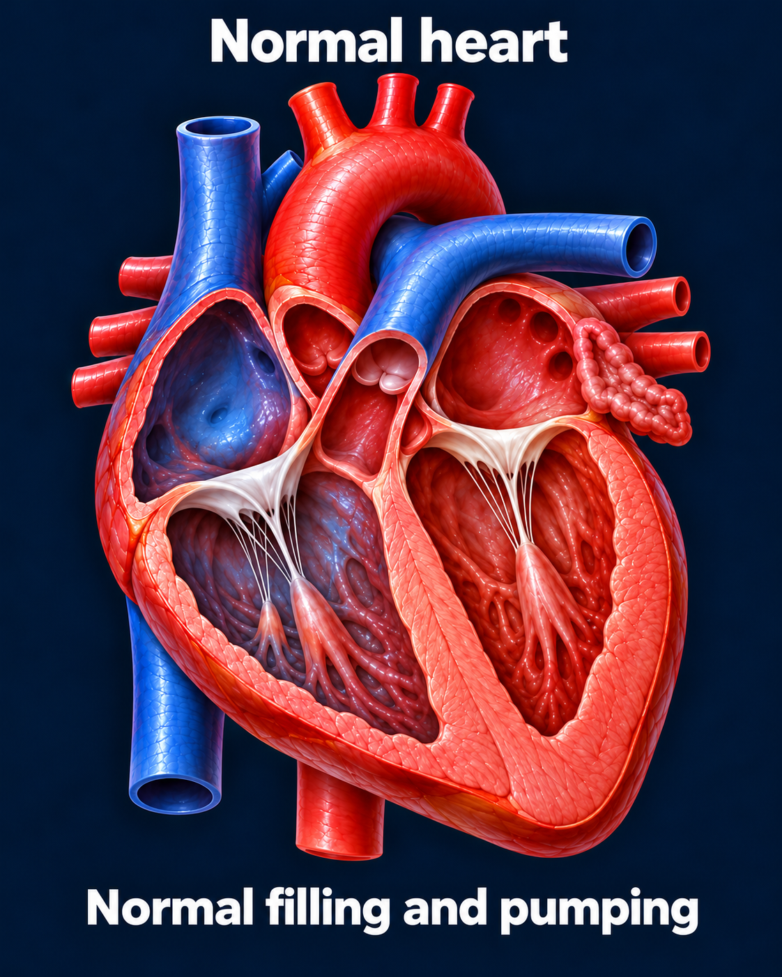
A healthy heart must fill and eject effectively. This is a simplified anatomical illustration.Open full-size image →
What does ejection fraction tell us?
Left ventricular EF is the proportion of blood in the ventricle that is ejected with each beat. If the LV fills with 100 mL and ejects 60 mL, the EF is 60%. EF helps classify heart failure and guide treatment, but does not describe every aspect of cardiac performance.
HFrEF · Reduced EF
LVEF: ≤40%
Meaning: Reduced ventricular ejection.
HFmrEF · Mildly reduced EF
LVEF: 41–49%
Meaning: Mildly reduced EF with supporting evidence of heart failure.
HFpEF · Preserved EF
LVEF: ≥50%
Meaning: Heart failure despite a preserved ejection fraction.
HFimpEF · Improved EF
LVEF: Previously ≤40%, now >40%
Meaning: EF has improved following a prior reduced-EF state.
Categories follow the AHA/ACC/HFSA framework. For EF >40%, evidence of increased filling pressures supports the diagnosis.[1]
HFrEF · Reduced pumping function
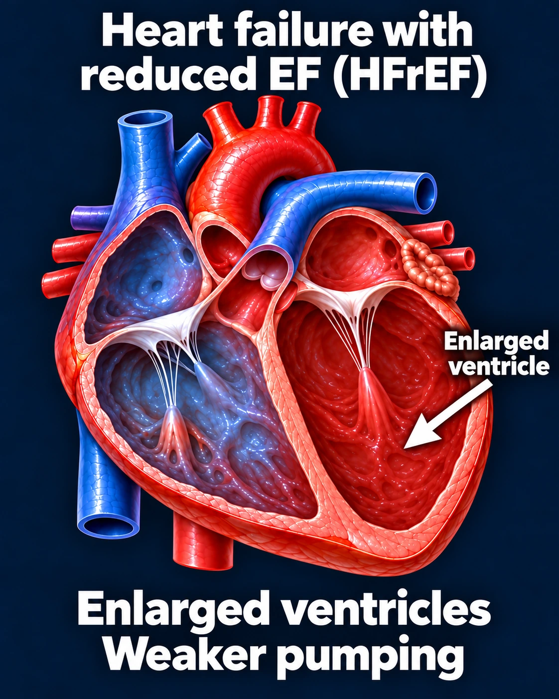
In HFrEF, the LV ejects a smaller fraction of its filled volume. The ventricle may enlarge, and pressure can rise behind it. Reduced output and congestion can coexist.[2]
A common HFrEF pattern: ventricular enlargement and reduced systolic function. Not every patient has this exact anatomy.Open full-size image →
HFpEF · Preserved EF, abnormal filling
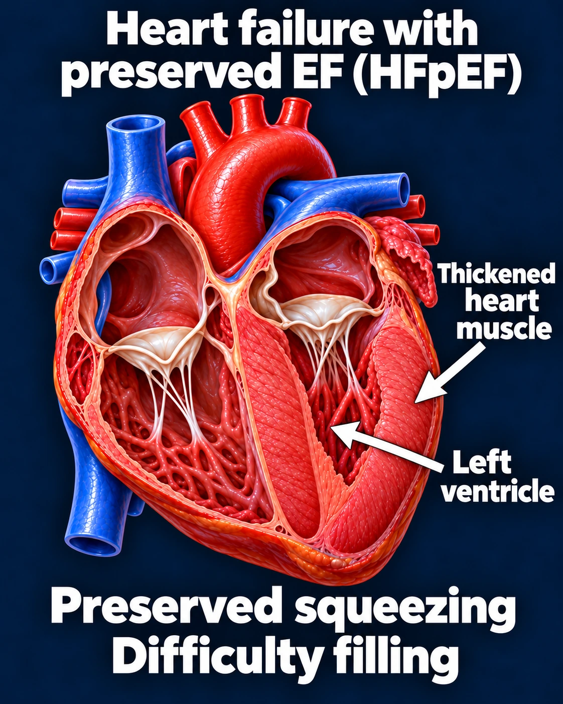
In HFpEF, abnormalities in relaxation, stiffness, and the cardiovascular system can raise filling pressures despite preserved EF. Pressures may rise particularly during exercise. A thickened LV is one possible pattern; it is not required in every patient.[2]
One HFpEF phenotype. Preserved EF does not establish normal filling, normal cardiac output, or the absence of heart failure. One example of HFpEF; not every patient has thickened heart muscle.Open full-size image →
Clinical distinction:
A patient with dyspnea and EF 60% may still have HFpEF. A patient with EF 60% and no supporting evidence of heart failure should not be labeled HFpEF solely because of that number.[1]
Congestion and poor perfusion produce different clues
Left-sided pressure → lungs
Exertional breathlessness, orthopnea, waking breathless at night, and pulmonary congestion.
Systemic venous congestion → body
Leg swelling, abdominal fullness/ascites, weight gain, and reduced appetite.
Reduced perfusion may cause fatigue, cool extremities, or impaired concentration. Findings often overlap, and edema alone is not specific for heart failure.[3]
Identify the underlying cause
Common contributors include ischemic disease/prior MI, hypertension, valve disease, cardiomyopathy, myocarditis, and persistent arrhythmias. Obesity, diabetes, kidney disease, and other comorbidities influence the clinical picture.
Cardiology evaluation identifies the cause and guides treatment. Correcting a contributing rhythm or valve problem can be important alongside heart failure therapy.[4]
Improved EF does not mean treatment is finished.
Patients with HFimpEF generally continue guideline-directed therapy to reduce relapse risk.[1]
Connect recognition to referral
New suspected heart failure, newly reduced EF, uncertain HFpEF, or persistent/worsening symptoms should prompt cardiology assessment. Severe resting breathlessness, hypoxemia, chest pain, or signs of shock require emergency evaluation.