Recognize the circuit, read the EKG, and understand why an organized rhythm still needs treatment and stroke-risk assessment.
UNDERSTAND THE RHYTHM
What is atrial flutter?
Atrial flutter is an organized electrical rhythm that makes the atria beat rapidly. Although the electrical activity is more organized than AFib, flutter can still cause significant symptoms, heart failure, and stroke.
An electrical signal repeatedly travels around a large circuit in the atrium—a mechanism called macroreentry.
The atria commonly activate around 250–350 times per minute, often near 300. The AV node blocks some of these signals from reaching the ventricles.
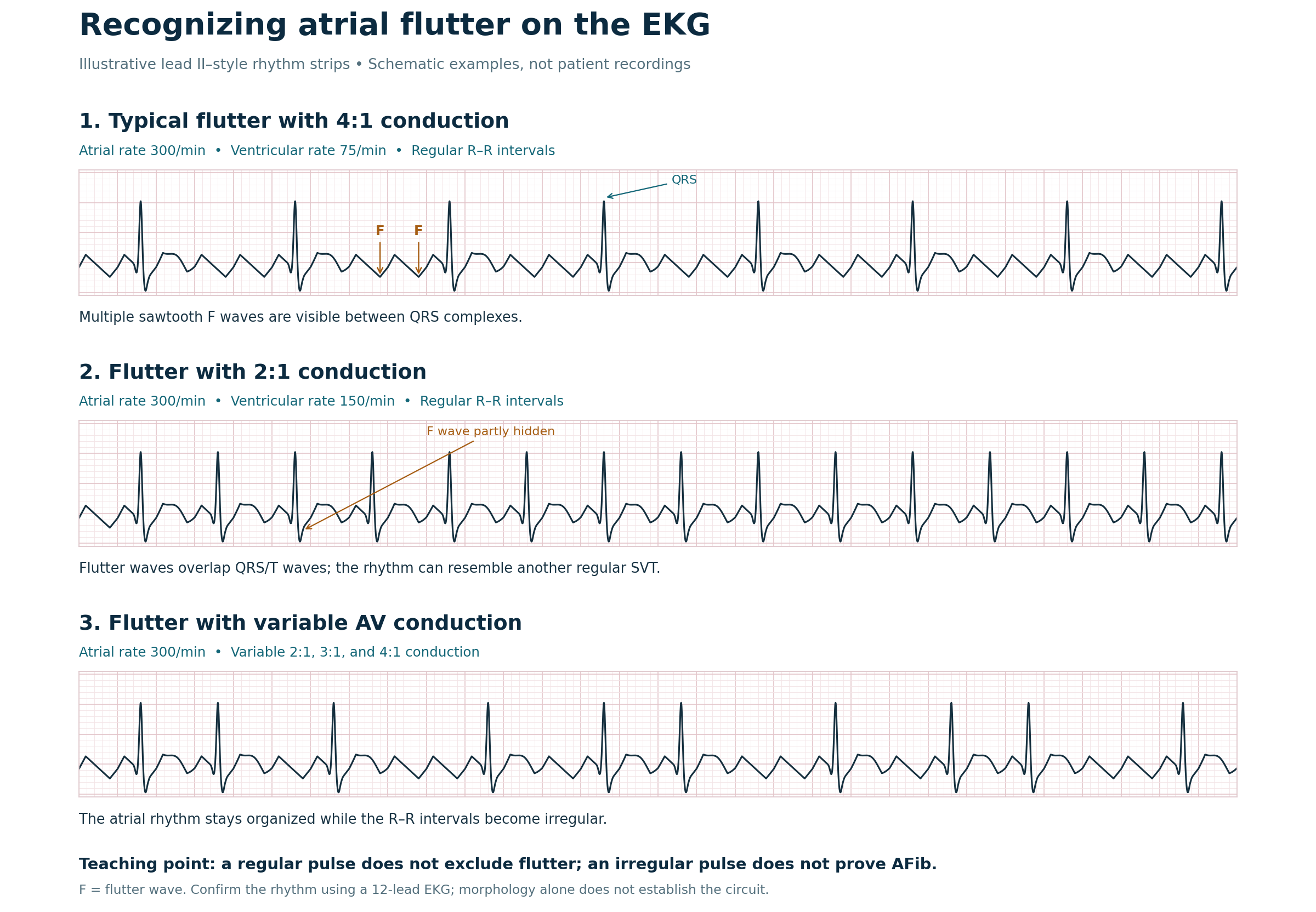
With 2:1 conduction, every second atrial signal reaches the ventricles. An atrial rate of 300 therefore produces a ventricular rate near 150 beats per minute.
Clinical clue
A persistent, regular narrow-complex tachycardia near 150 should raise suspicion for flutter with 2:1 conduction. The rate alone does not establish the diagnosis.
KNOW THE CIRCUIT
Typical versus atypical flutter
Typical atrial flutter
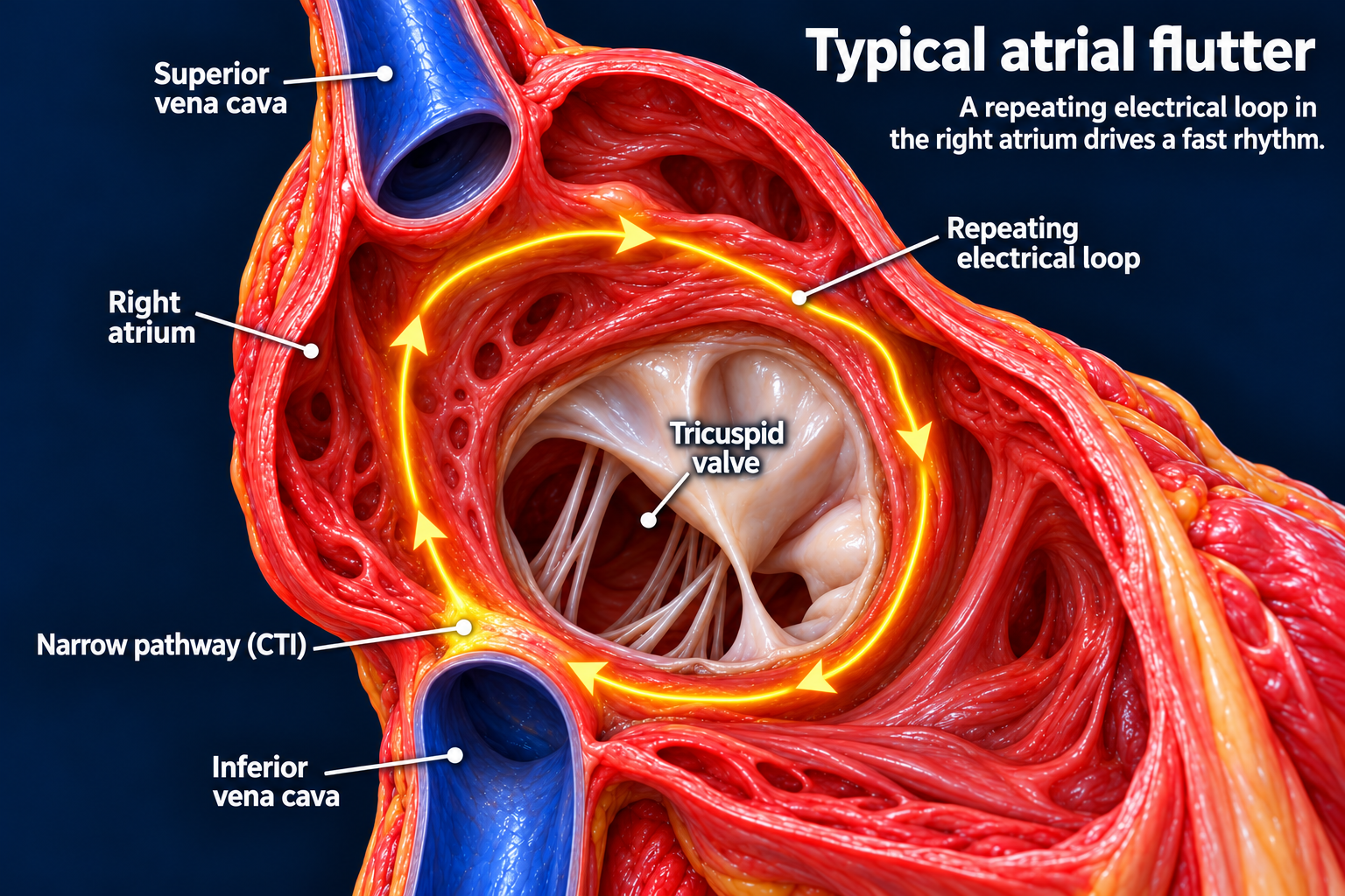
The right atrial circuit passes through the CTI, between the tricuspid annulus and inferior vena cava. Arrow direction is illustrative; typical flutter can rotate in either direction.Open full-size image →
Typical flutter travels around the tricuspid annulus in the right atrium and depends on the cavotricuspid isthmus (CTI). It may occur independently or alongside AFib. An ablation line across the CTI interrupts this circuit.
Atypical atrial flutter
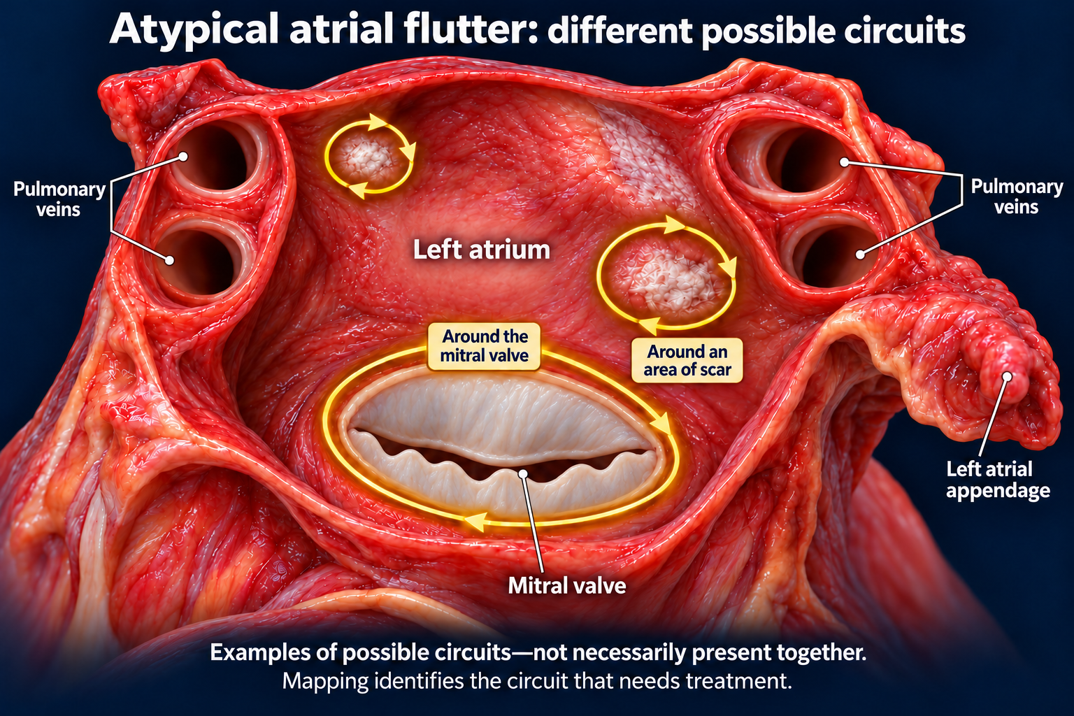
These illustrate alternative left atrial circuits, not necessarily rhythms occurring together. Atypical flutter can also arise in the right atrium.Open full-size image →
Atypical flutter does not depend on the CTI. It uses a different circuit in either atrium, often involving scar. It may occur after atrial surgery or AFib ablation, or with underlying atrial disease. Mapping identifies the circuit and the pathway that needs treatment.
Typical flutter has a predictable circuit.
Atypical flutter requires a more individualized mapping approach.
READ THE EKG
Recognizing flutter on the EKG
Look for repetitive atrial activity rather than normal, discrete P waves.
Common counterclockwise typical flutter often produces negative sawtooth waves in leads II, III, and aVF, with positive activity in V1.
Clockwise typical flutter can have a different appearance.
With 2:1 conduction, flutter waves may be hidden within the QRS complexes or T waves.
Fixed conduction can produce a regular ventricular rhythm; variable conduction can make it irregular.
Schematic teaching examples—not patient recordings. Open the image for a larger view. Atrial rate is 300/min throughout; ventricular timing changes with AV conduction.
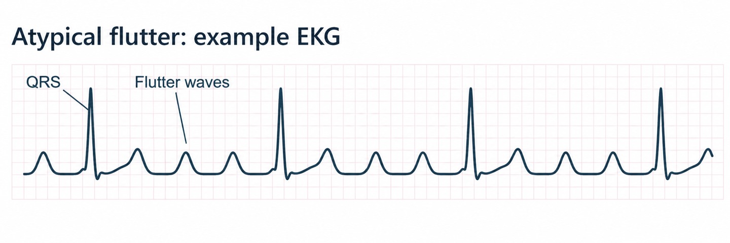
An atypical flutter EKG illustration
Rounded atrial waves are one possible appearance. This schematic does not establish the location or mechanism of the circuit.Open full-size image →
An unusual EKG appearance does not automatically mean atypical flutter.
The circuit determines the classification, and EP mapping may be needed to confirm it.
LOOK BEYOND THE PULSE
Why does flutter matter?
Patients may experience palpitations, fatigue, shortness of breath, reduced exercise tolerance, dizziness, or chest discomfort. Some have few symptoms.
A sustained rapid ventricular rate can impair ventricular function and cause tachycardia-mediated cardiomyopathy. A patient who feels relatively well may still need treatment.
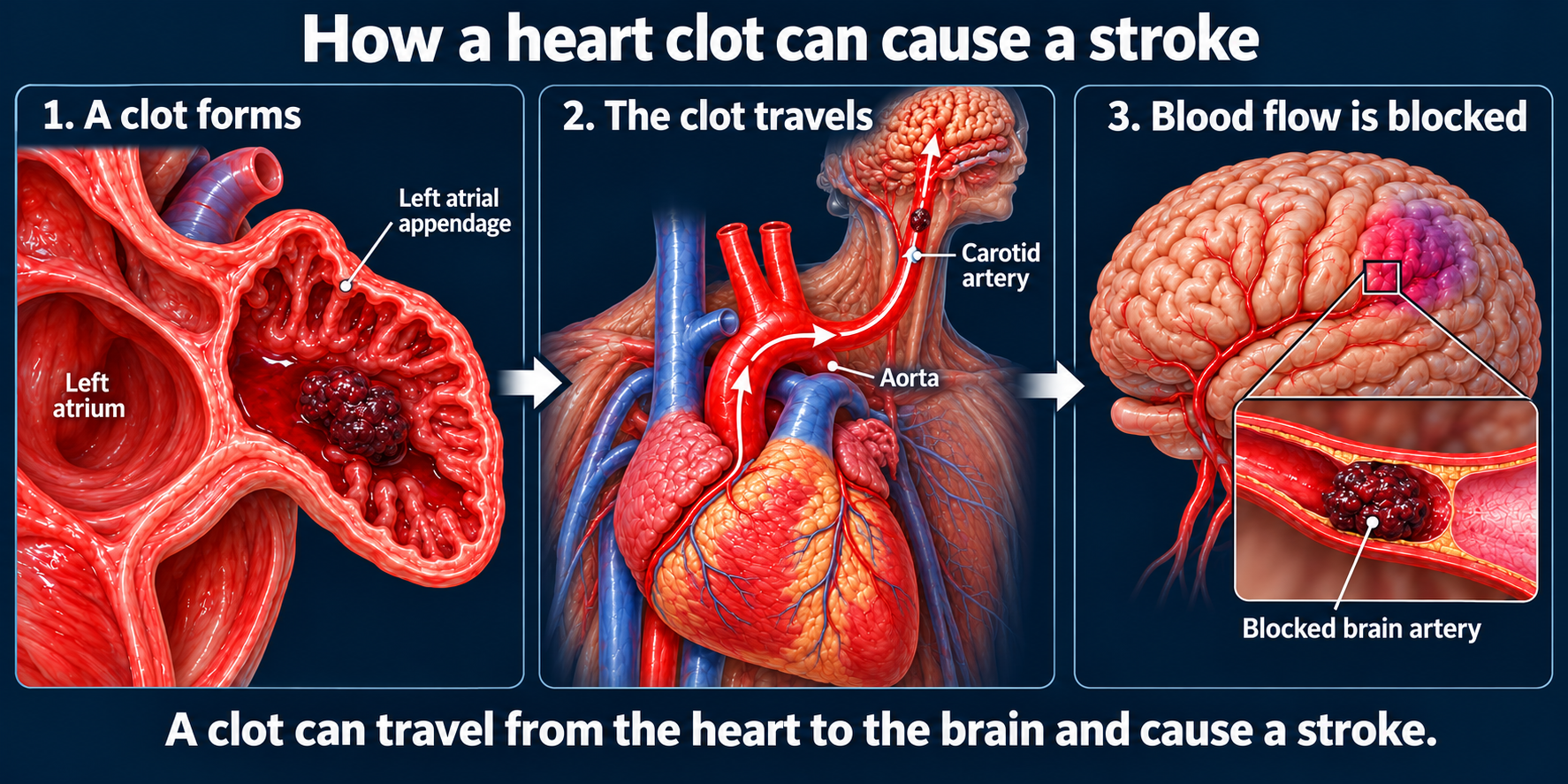
Atrial flutter carries thromboembolic risk. Assess anticoagulation using the same stroke-risk framework used for AFib.
An organized rhythm or regular pulse does not remove the need to assess stroke risk. Flutter and AFib frequently coexist, and AFib may appear after successful flutter ablation.
Successful flutter ablation does not automatically mean anticoagulation can be stopped. That decision requires review of stroke risk, AFib history, and follow-up monitoring.
CONNECT LEARNING TO REFERRAL
Why refer to EP?
For typical flutter, ablation can interrupt the defined circuit and provide durable control of that rhythm. Atypical flutter can also be treated, although the procedure is usually more complex.
Offer EP evaluation early to discuss the mechanism and treatment options. Eliminating a flutter circuit does not eliminate a patient's separate risk of developing AFib.