Identify red flags and understand the diagnostic sequence.
Assess symptoms and urgency
New severe dyspnea, shock, syncope with concerning cardiac findings, sustained arrhythmia, or rapidly worsening HF requires urgent assessment. For stable patients, review symptoms, volume status, EKG, echo, exposures, cancer therapy, and a three-generation family history where feasible.
Coordinate echo and selected cardiac MRI to define function, distribution of thickening, scar, or inflammation. Evaluate ischemia and reversible contributors where indicated. Family screening and genetic counseling are important when an inherited disorder is suspected.
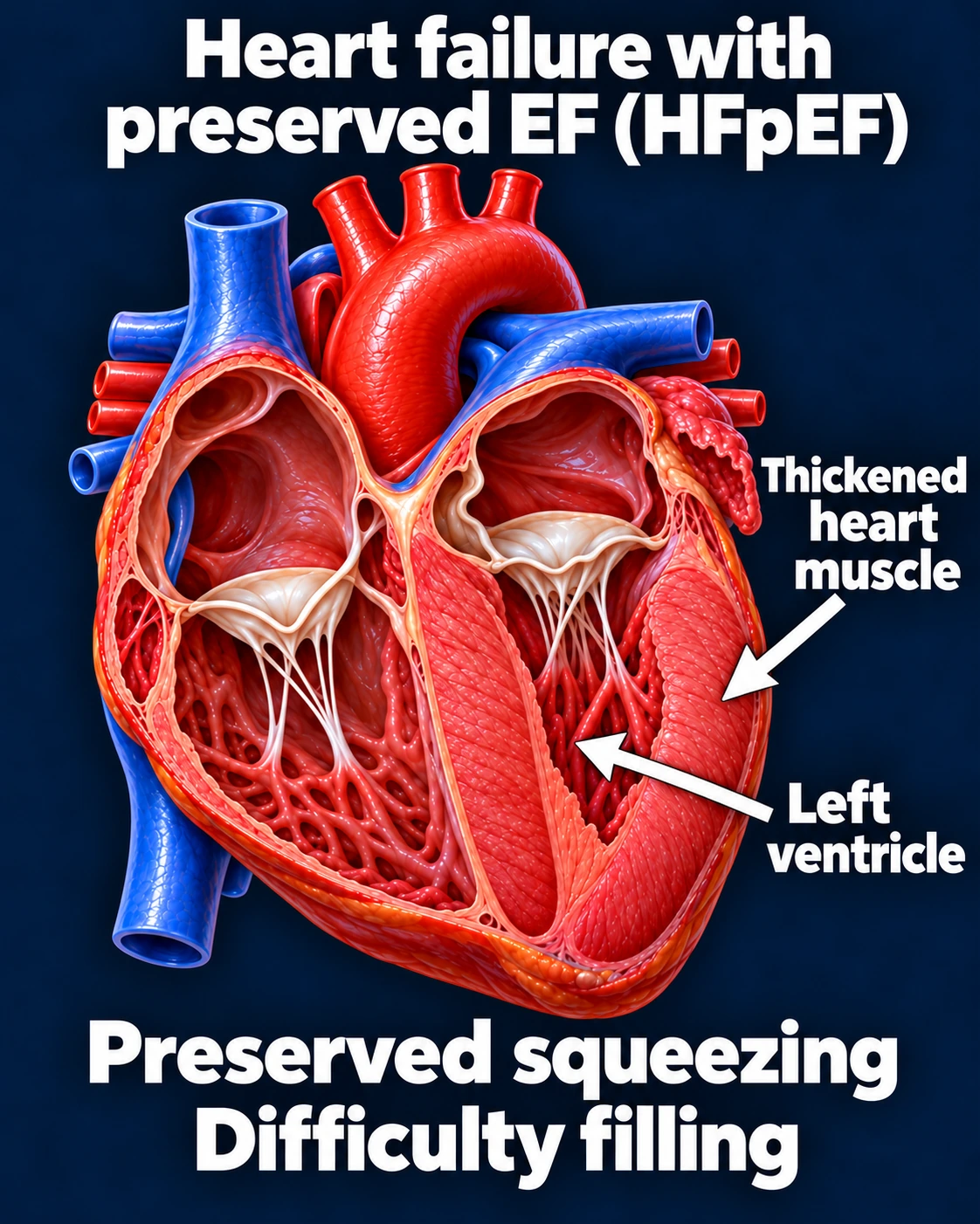
An illustration of a filling problem. This is not an amyloid-specific image or diagnostic pattern. One example of HFpEF; not every patient has thickened heart muscle.Open full-size image →[1][3]
Do not skip the AL screen
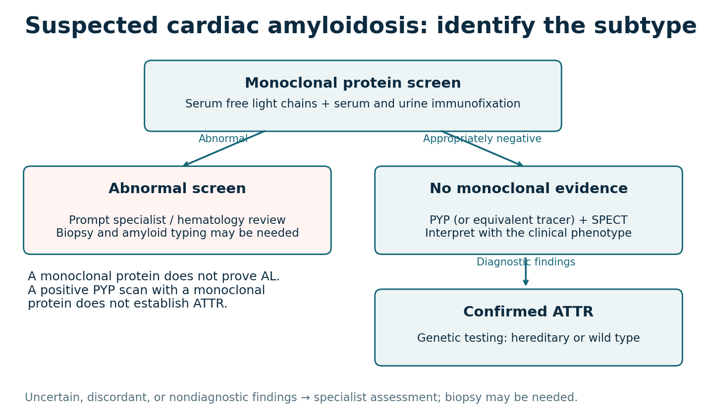
Clinical sequence for suspected cardiac amyloidosis. Specialist interpretation and tissue typing may be needed.Open full-size image →
When amyloidosis is suspected, the monoclonal protein screen includes serum free light chains plus serum and urine immunofixation. Routine protein electrophoresis alone is insufficient. Interpret free-light-chain results in the setting of kidney function.
Abnormal results require prompt specialist review; a monoclonal protein does not by itself prove AL. Tissue confirmation and amyloid typing may be needed. A negative fat-pad biopsy does not exclude cardiac amyloidosis.
After appropriate exclusion of a monoclonal process, characteristic technetium-based cardiac uptake with confirmatory SPECT imaging can support a nonbiopsy ATTR diagnosis in the proper context. A positive PYP scan with a monoclonal protein cannot simply be labeled ATTR; biopsy and typing may be required.
CMR supports suspicion but does not reliably type AL versus ATTR. Confirmed ATTR warrants genetic testing to distinguish hereditary from wild-type disease.