Understand echo, stress imaging, CT, MRI, and rhythm monitoring.
Echocardiography: the structure and function test
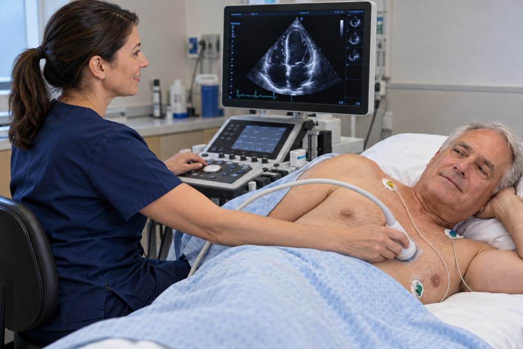
Echo assesses chamber size, systolic/diastolic function, valves, and pericardial findings. LVEF describes the fraction of LV blood volume ejected per beat; it is not the percentage of the heart that works.
Ultrasound images assess structure and function. Resting echo does not directly exclude coronary stenosis.Open full-size image →
TEE provides selected higher-detail views, including the LAA and prosthetic valves, with different preparation and procedural considerations.
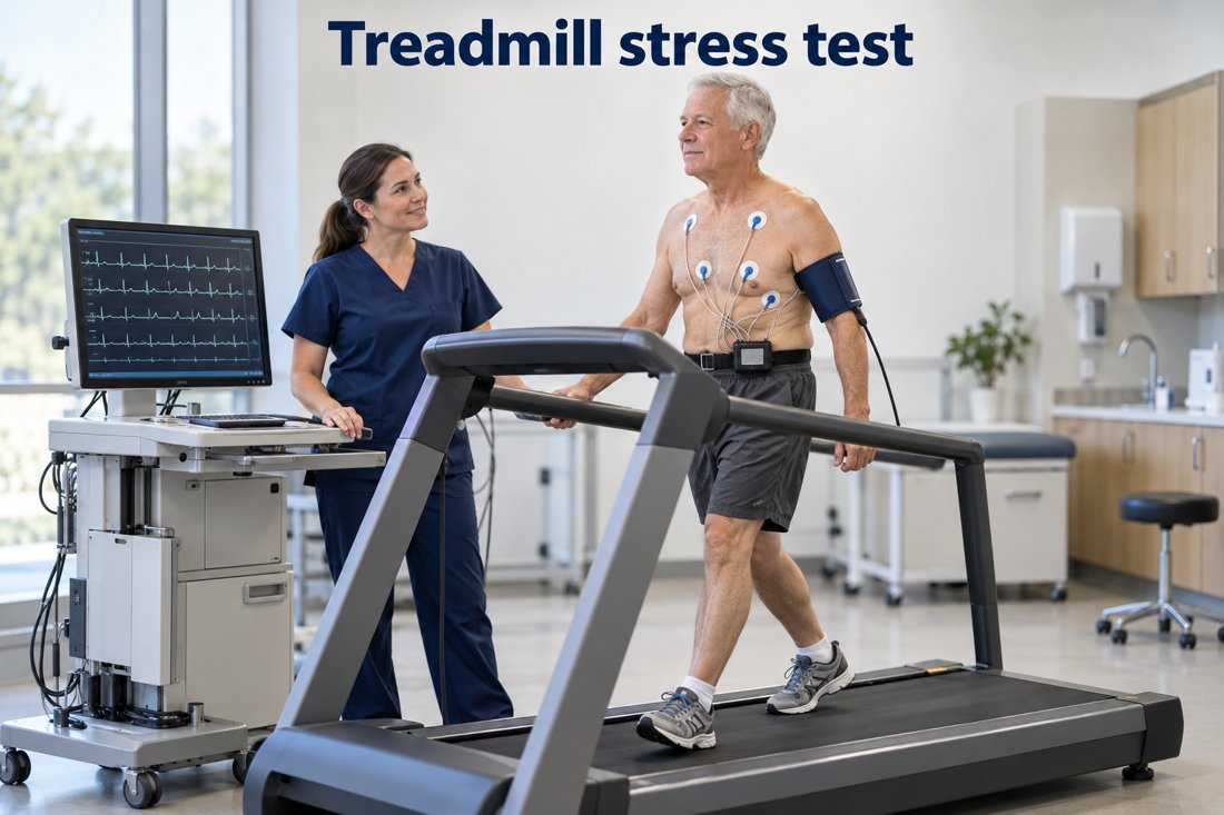
Exercise testing adds information on symptoms, functional capacity, BP, and rhythm. Stress echo evaluates induced wall-motion changes; SPECT or PET evaluates perfusion. Pharmacologic stress is used when exercise is unsuitable.
Choose the test based on exercise ability, baseline EKG, clinical question, and local expertise.Open full-size image →
PET may quantify myocardial blood flow and flow reserve. Interpret defects alongside artifacts and the report’s quality assessment. Do not treat an automated summary as the final interpretation.
CCTA uses contrast to depict coronary anatomy. CAC is a noncontrast measure of calcified plaque for selected prevention decisions; it is not a replacement for evaluating symptomatic coronary disease.
Cardiac MRI characterizes myocardium, scar, and function. Before ordering, assess contrast considerations, kidney function when relevant, claustrophobia, and any implanted device. Device presence requires a dedicated MRI safety pathway rather than a blanket assumption.
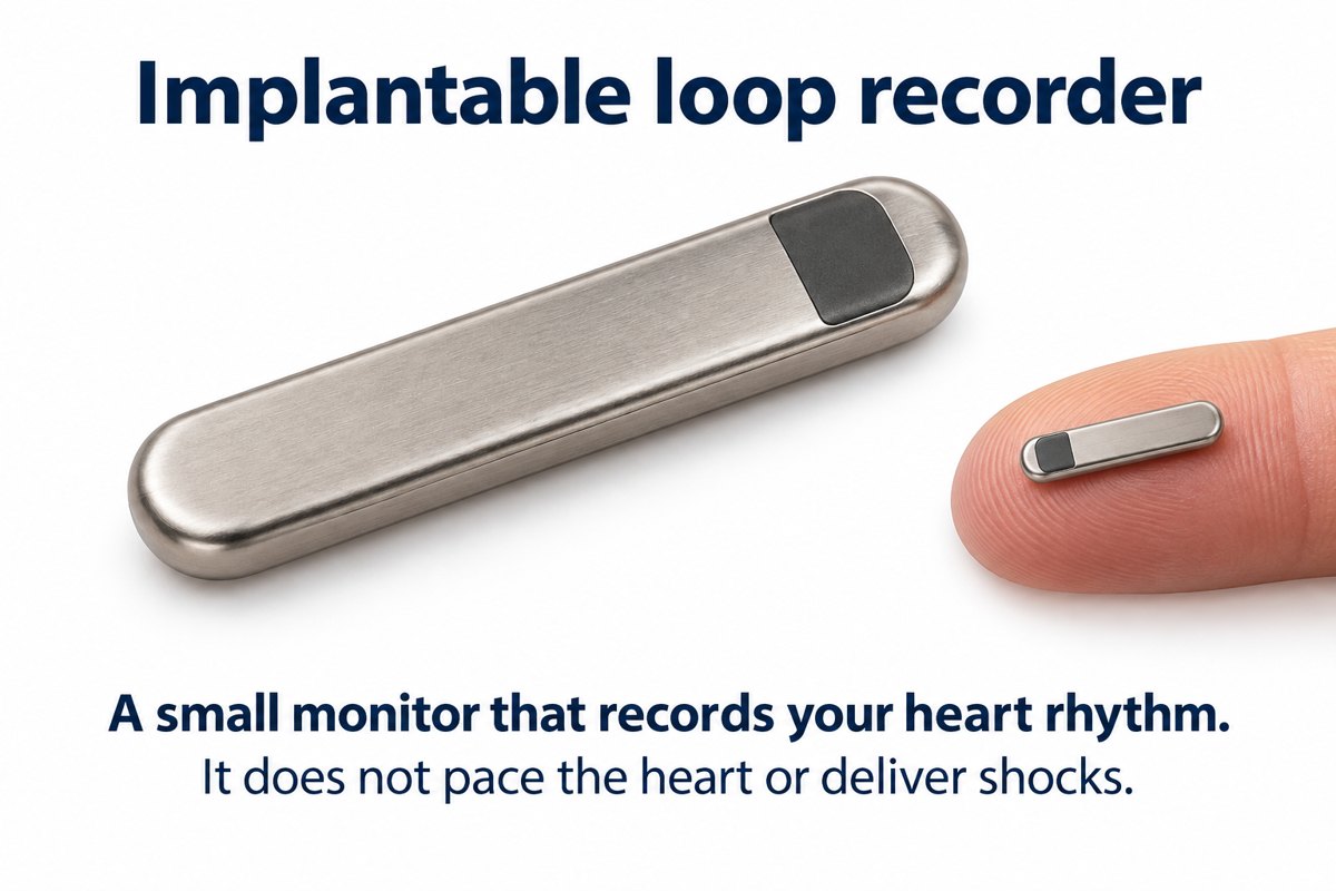
Match recording duration to symptom frequency: brief Holter for frequent events, extended patch/event monitoring for less frequent symptoms, and selected implantable loop recording for infrequent unexplained events. Record symptom timing and review actual strips.
A diagnostic rhythm recorder. It does not pace the heart or deliver a shock.Open full-size image →[6]