Understanding AFib. Recognize the disease behind the rhythm.
AFib is a progressive atrial disease. Understand its triggers, substrate, and consequences so the first encounter leads to a long-term plan.
UNDERSTAND THE RHYTHM
What is atrial fibrillation?
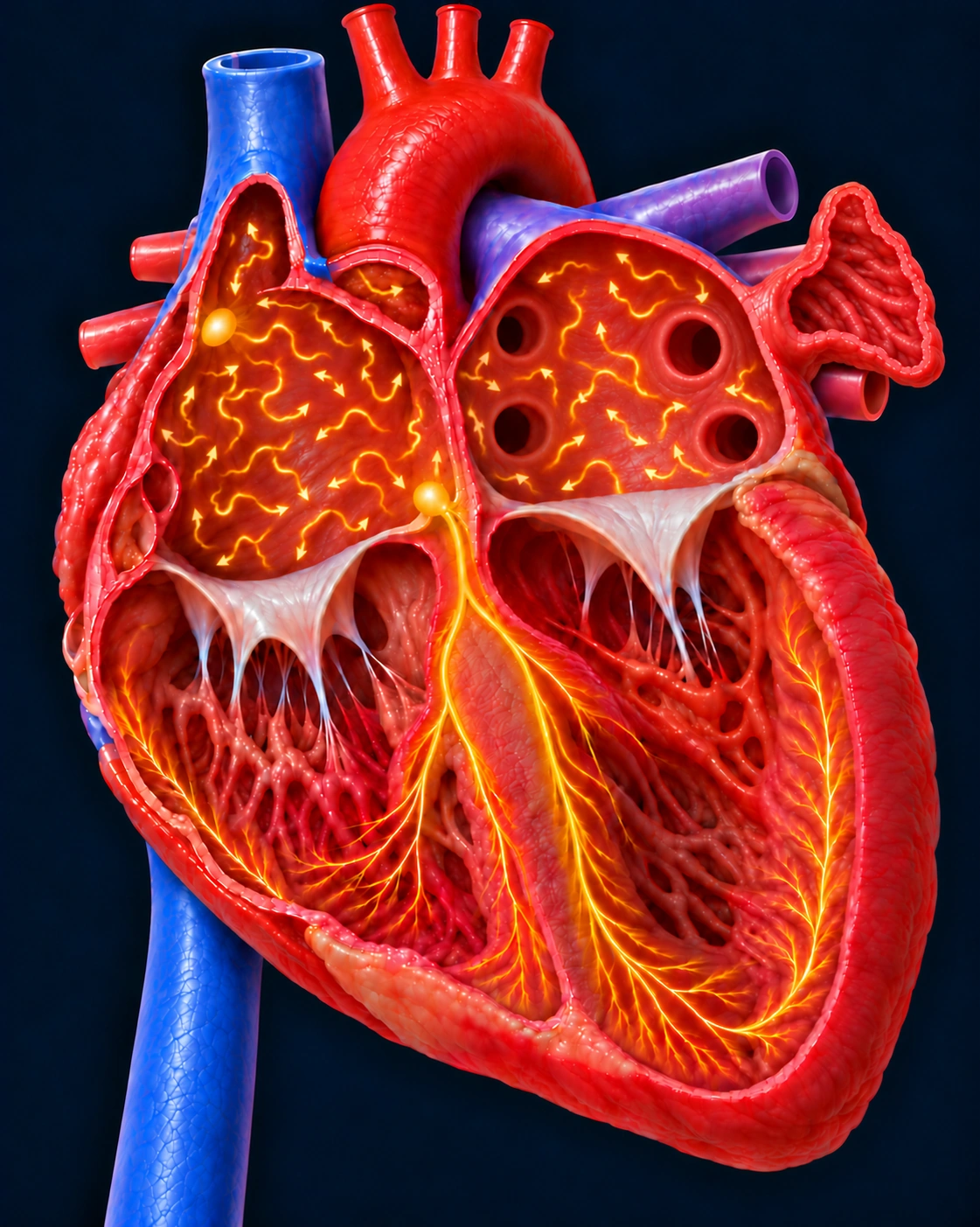
AFib is disorganized electrical activation of the atria. Instead of contracting together, the upper chambers fibrillate, reducing effective atrial contraction.
The AV node conducts some of these signals to the ventricles, producing an irregular ventricular rhythm that may be rapid. A controlled ventricular rate does not mean the atria have returned to normal rhythm.
Follow one normal heartbeat
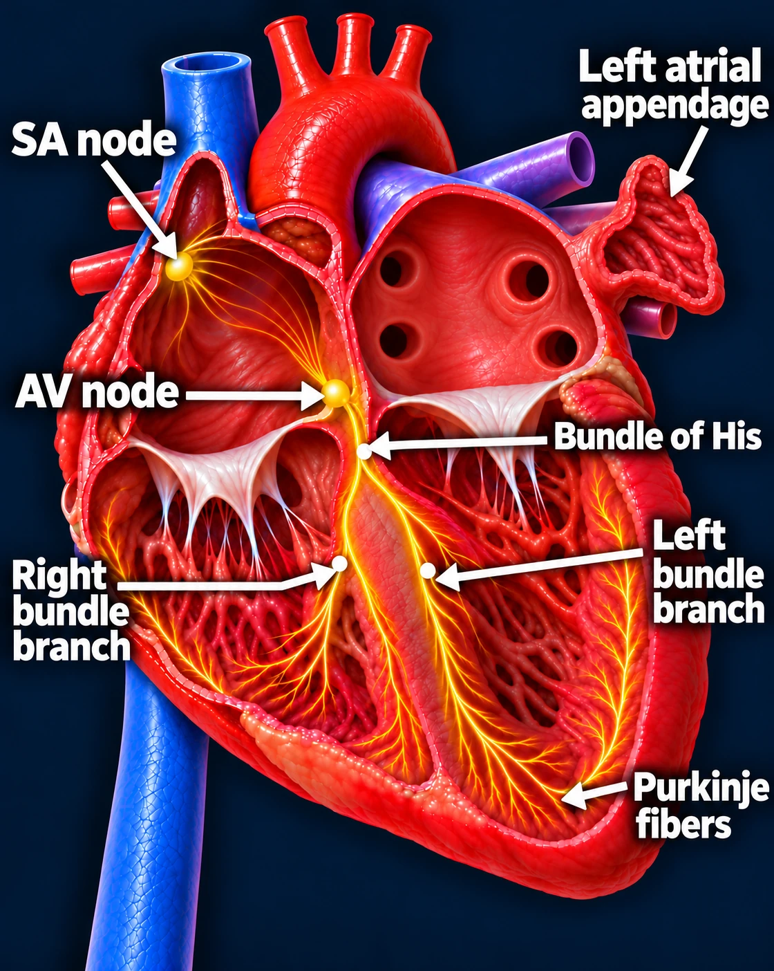
SA node → Atria → AV node → Ventricles
The SA node starts the signal. The heart’s natural pacemaker in the right upper chamber begins each normal sinus beat.
The signal spreads through both atria. The upper chambers contract, moving blood into the ventricles.
The AV node briefly delays the signal. This pause gives the ventricles time to finish filling.
The signal activates the ventricles. It travels through the bundle of His, right and left bundle branches, and Purkinje fibers. The lower chambers contract, pumping blood to the lungs and body.
The atria contract first; the ventricles follow. This organized sequence produces a coordinated heartbeat.
Regular R–R intervals and consistent P waves before each QRS.
Atrial fibrillation
Irregularly irregular R–R intervals without consistent P waves. Both strips are schematic, not patient recordings.
Symptoms may be obvious—or absent
Patients may report palpitations, fatigue, reduced exercise tolerance, dyspnea, dizziness, or chest discomfort. Some have few symptoms despite a meaningful AFib burden. Symptom severity alone does not establish stroke risk or disease stage.
TRIGGERS & SUBSTRATE
How does AFib start?
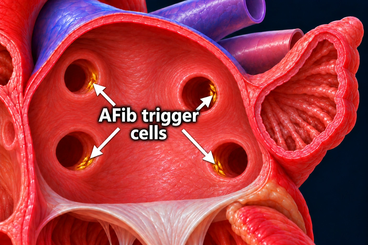
Premature atrial impulses commonly arise from myocardial sleeves extending into the pulmonary veins. These triggers can initiate AFib when the atria are susceptible to sustaining disorganized activation.
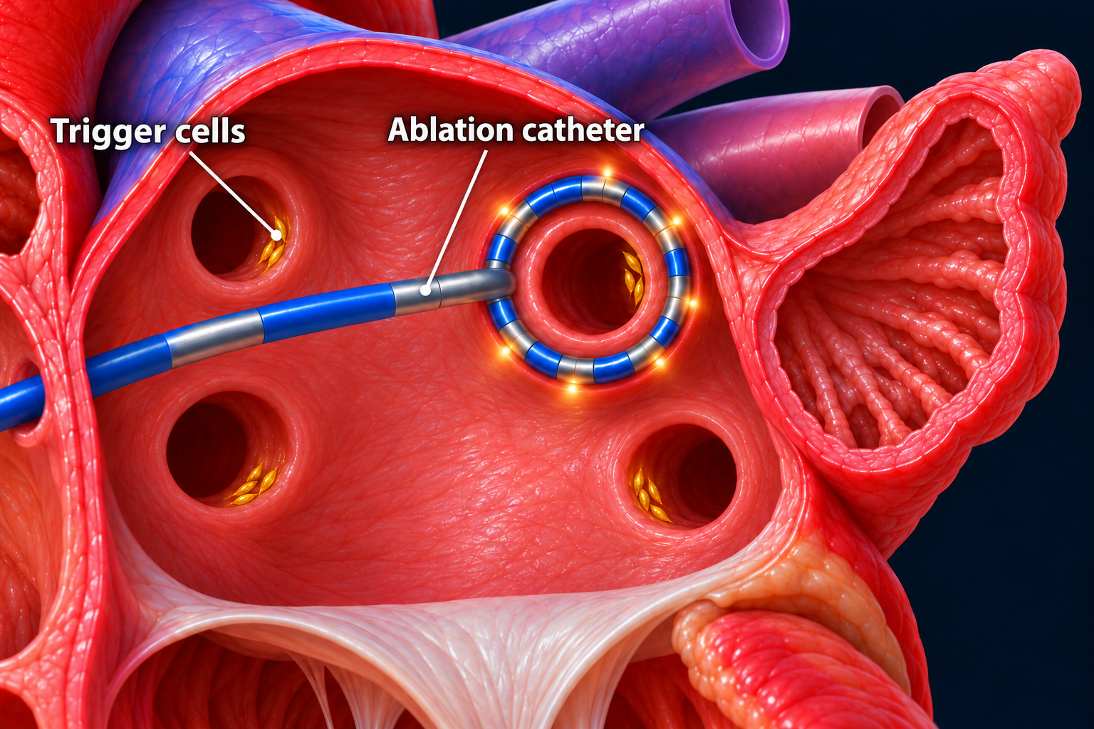
The labeled gold clusters represent potential AFib trigger cells in muscle sleeves near the pulmonary vein openings. Extra electrical signals from these sites can start an AFib episode. Cell size and location are simplified for teaching.Open larger image (new tab)
A trigger starts the episode; the atrial substrate helps sustain it. Fibrosis, atrial enlargement, conduction abnormalities, and electrical remodeling can make AFib more persistent. Hypertension, obesity, sleep apnea, alcohol use, heart failure, aging, and other factors can contribute to this environment.
AFIB IS A PROGRESSIVE DISEASE
AFib can create conditions for more AFib
Repeated or sustained AFib can promote electrical and structural remodeling. Over time, episodes may become longer, more frequent, and harder to suppress.
The course varies between patients; progression is not inevitable at a fixed pace. But a quiet symptom history or controlled pulse does not establish that the underlying disease is stable.
Electrophysiologists: the cardiac firefighters
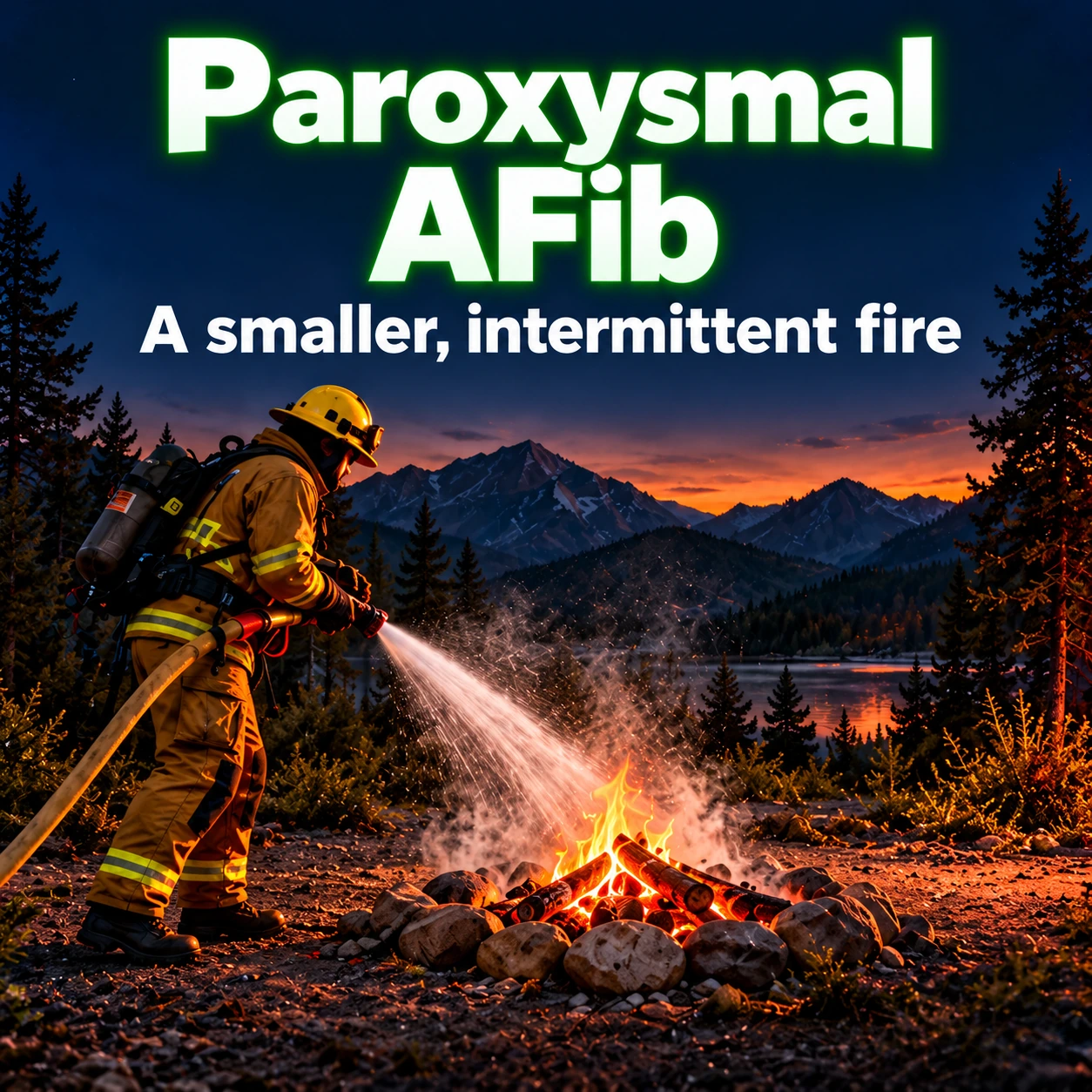
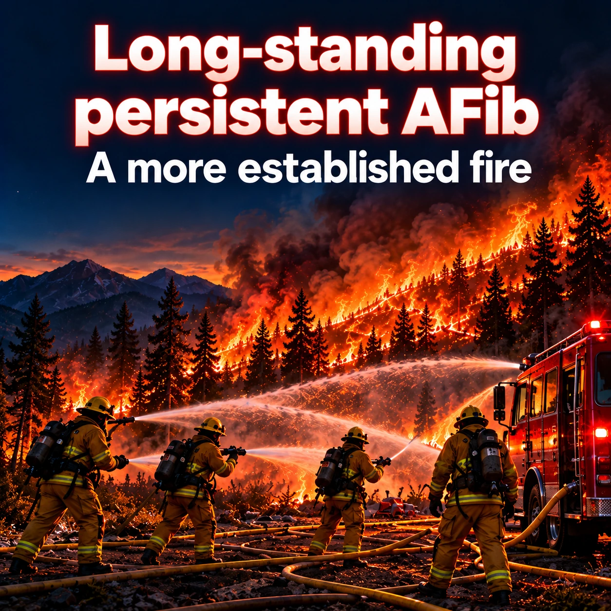
Paroxysmal AFibA smaller, intermittent fire. Triggers may play a larger role, and earlier rhythm control offers a better opportunity to maintain sinus rhythm.Open full-size image →Persistent AFibA more sustained fire with a greater substrate contribution. Rhythm control can still improve symptoms and reduce AFib burden.Open full-size image →Long-standing persistent AFibA more established fire. Maintaining sinus rhythm is often harder and may require more than one treatment, but an EP assessment can still identify meaningful options.Open full-size image →
Think of trigger cells as sparks, AFib episodes as the fire, and atrial remodeling as the coals and fuel that make future episodes easier to sustain. EPs try to suppress the fire and isolate the sparks before a large bed of coals develops.
Paroxysmal AFib
A smaller, intermittent fire. Triggers may play a larger role, and earlier rhythm control offers a better opportunity to maintain sinus rhythm.
Persistent AFib
A more sustained fire with a greater substrate contribution. Rhythm control can still improve symptoms and reduce AFib burden.
Long-standing persistent AFib
A more established fire. Maintaining sinus rhythm is often harder and may require more than one treatment, but an EP assessment can still identify meaningful options.
Permanent AFib is a shared treatment decision, not the inevitable next stage. Pattern definitions and documentation belong in Section 2.
Earlier rhythm-control discussions matter.
Early rhythm control can improve outcomes in appropriately selected patients. Catheter ablation can reduce recurrence and progression in selected patients. Do not wait for years of AFib or repeated medication failure before offering EP evaluation.
TREAT THE SOURCE
What does AFib ablation do?
Pulmonary vein isolation (PVI) creates an electrical barrier between the pulmonary-vein myocardial sleeves and the left atrium.
1. How the catheter reaches the left atrium
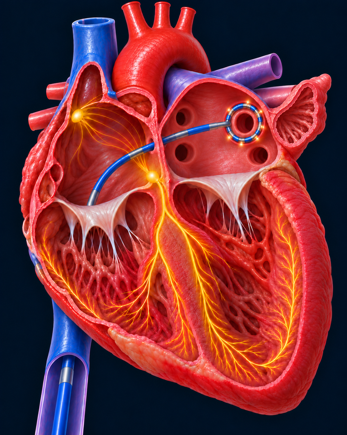
The catheter travels from a vein in the groin through the inferior vena cava (IVC) to the right atrium, then crosses the wall between the upper chambers to reach the left atrium. Its circular tip is shown around one pulmonary vein opening. Pulmonary vein isolation (PVI) creates an electrical barrier that keeps signals from the pulmonary veins from entering the atrium. All pulmonary veins are treated during PVI. This is a simplified illustration of the route and catheter position.Open larger image (new tab)
2. How ablation isolates the trigger cells
The circular ablation catheter is shown around a pulmonary vein opening. The gold clusters represent AFib trigger cells. Ablation creates an electrical barrier that keeps signals from the pulmonary veins from entering the left atrium.Open larger image (new tab)
Signals arising inside an electrically isolated vein cannot conduct into the atrium through the blocked pathway. PVI is the foundation of AFib ablation. Additional treatment may be considered according to the patient's rhythm and atrial substrate.
Pulsed field ablation (PFA) uses brief electrical pulses to produce irreversible electroporation; radiofrequency and cryoablation use thermal energy. The therapeutic goal is durable electrical isolation.
Ablation generally maintains rhythm more effectively than antiarrhythmic medication in appropriately selected patients. It can reduce AFib burden and improve quality of life, but recurrence, medication use, or repeat ablation may still occur.
Suppression and long-term control—not a guaranteed lifelong cure.
AFib requires ongoing follow-up, risk-factor treatment, and reassessment of stroke prevention even after successful ablation.
PROTECT THE BRAIN
Why does AFib increase stroke risk?
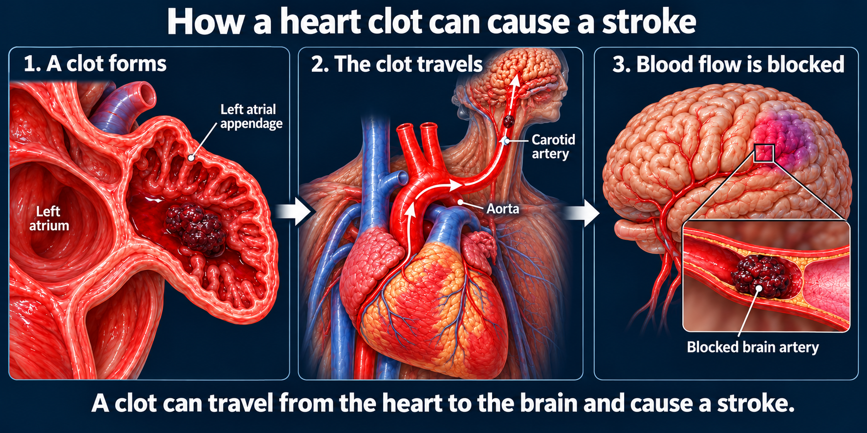
Ineffective atrial contraction can permit blood stasis in the left atrial appendage (LAA), where thrombus may form. A clot can then embolize to a cerebral artery and cause ischemic stroke.
AFib is associated with about a fivefold increase in stroke risk at the population level. An individual patient's absolute risk depends on clinical risk factors; the practical assessment and anticoagulation decisions are covered in Section 2.
Stroke prevention and rhythm control solve different problems. Restoring sinus rhythm does not automatically remove the indication for anticoagulation.
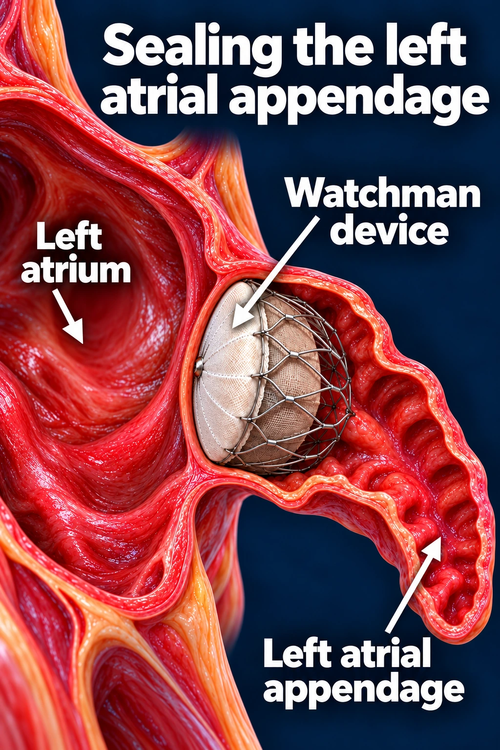
Where does Watchman fit?
Watchman closes the LAA to reduce embolization of clots formed there.Open full-size image →
Left atrial appendage occlusion is an option for selected patients with AFib and elevated stroke risk who cannot take long-term anticoagulation, or who have a high bleeding risk after individualized assessment. It does not treat the AFib rhythm or replace anticoagulation needed for another condition.
Implantation carries procedural risks. Short-term antithrombotic treatment and follow-up imaging are usually required, according to the device and clinical plan.
THE CLINICAL TAKEAWAY
Offer EP evaluation early—even after the first episode
AFib is more than a fast pulse. It is associated with stroke, heart failure, hospitalization, and reduced quality of life. It is also associated with cognitive decline; that association does not establish that rhythm treatment prevents dementia.
Our referral approach is to offer every patient with documented AFib an EP consultation to understand the disease and compare long-term options. The consultation does not mean every patient needs an ablation.
Earlier referral preserves time for an informed rhythm-control decision. Persistent and long-standing persistent AFib still deserve assessment rather than being dismissed as “too late.”