Understand each device, its purpose, and the main reasons for implantation.
Start with the purpose
Cardiac devices provide different forms of electrical treatment. Knowing whether the patient needs bradycardia support, protection from ventricular arrhythmias, or ventricular resynchronization helps frame the referral.
Pacemaker
Main purpose: Supports a slow or unreliable rhythm.
Defibrillation shocks?: No
ICD
Main purpose: Detects and treats dangerous ventricular arrhythmias.
Defibrillation shocks?: Yes
CRT-P
Main purpose: Coordinates ventricular activation, with pacing support.
Defibrillation shocks?: No
CRT-D
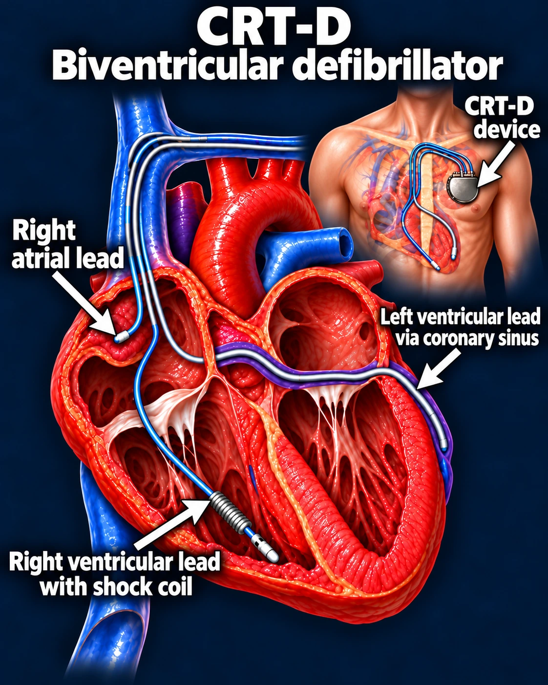
Main purpose: Combines resynchronization and ICD protection.
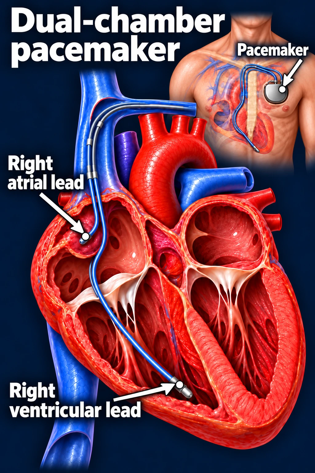
A pacemaker senses electrical activity and delivers small impulses when needed. A dual-chamber system can sense or pace both the right atrium and ventricle, coordinating their timing.[1]
Typical indications include symptomatic sinus node dysfunction and significant AV block. Acquired Mobitz II, high-grade, or complete AV block without a reversible or physiologic cause can warrant permanent pacing even without symptoms.[4]
Conventional dual-chamber pacemaker. The illustration shows an RV apical lead; actual pacing location is individualized.Open full-size image →
The pacing indication and the pacing strategy are separate decisions.
Once pacing is needed, EP selects chamber support, lead position, and programming according to the patient’s conduction, ventricular function, and expected pacing burden.
ICDs · Protect against dangerous ventricular rhythms
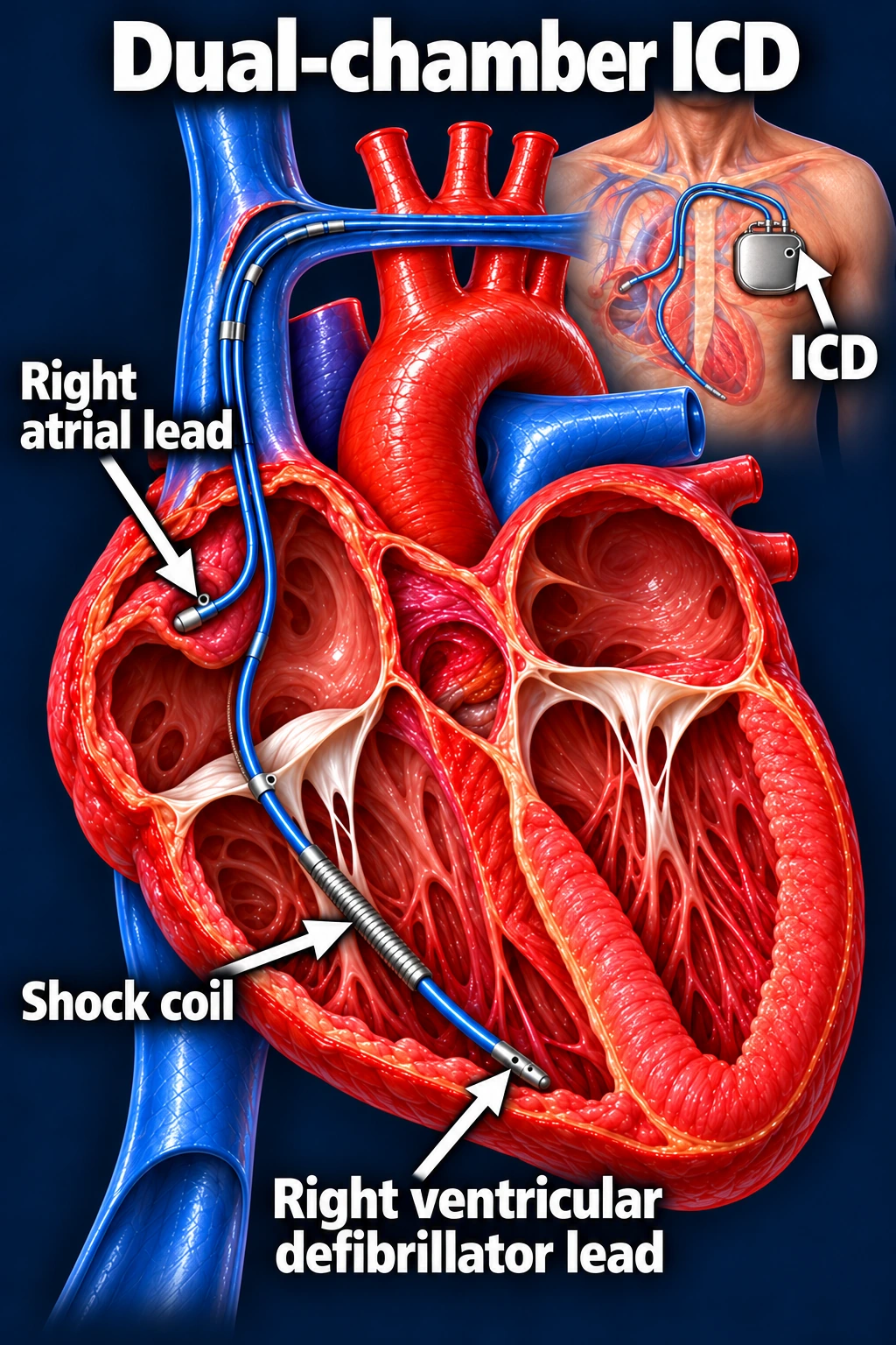
An implantable cardioverter-defibrillator monitors for ventricular tachycardia (VT) and ventricular fibrillation (VF). Depending on the rhythm and programming, a transvenous ICD may deliver antitachycardia pacing (ATP) to interrupt VT or a shock to terminate a dangerous arrhythmia. Transvenous ICDs can also provide bradycardia pacing.[2], [5]
The shock coil identifies a transvenous defibrillation lead. Not all ICDs require an atrial lead.Open full-size image →
Secondary prevention
For selected survivors of VT/VF arrest or patients with sustained VT not due to a reversible cause, when meaningful survival beyond one year is expected.[5]
Primary prevention
Before an arrest occurs, selected patients with persistently reduced EF despite appropriate therapy may benefit. A common indication is LVEF ≤35% with NYHA II–III symptoms on chronic guideline-directed therapy, appropriate timing after MI/revascularization, and expected meaningful survival beyond one year.[3]
An ICD indication is not determined by EF alone. Etiology, treatment duration, recovery potential, arrhythmic history, comorbidities, and preferences matter. Some inherited or arrhythmogenic disorders have separate risk-based indications.
A subcutaneous ICD avoids a transvenous lead but does not provide ongoing bradycardia pacing, ATP, or CRT. Those needs influence system selection.[5]
CRT · Coordinate ventricular activation
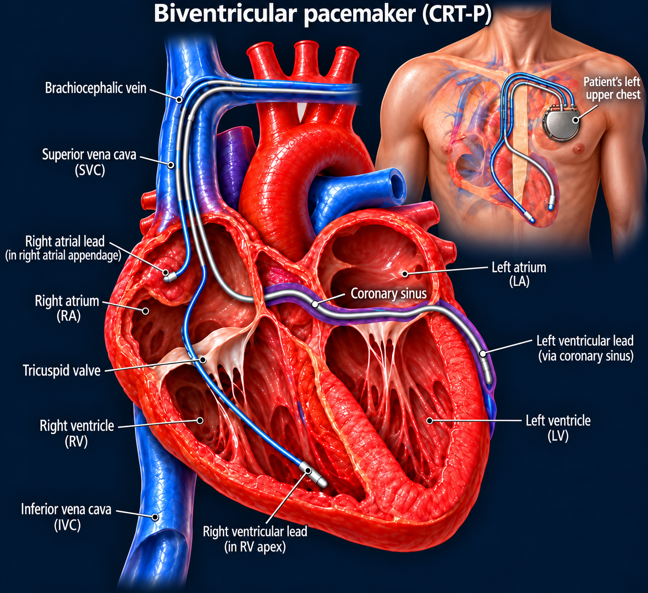
Cardiac resynchronization therapy addresses electrical dyssynchrony. Conventional biventricular CRT paces the right ventricle and the left ventricular surface through a coronary sinus branch.
A strong guideline-supported indication is LVEF ≤35%, sinus rhythm, LBBB with QRS ≥150 ms, and NYHA II, III, or ambulatory IV symptoms despite guideline-directed therapy. In appropriately selected patients, CRT can improve symptoms and ventricular function and reduce heart failure hospitalization and mortality. Other QRS patterns and pacing-related situations require individualized assessment.[3]
CRT-P provides resynchronization pacing. The LV lead sits in a coronary vein on the heart’s surface, not inside the LV cavity.Open full-size image →CRT-D adds defibrillation capability to resynchronization. CRT-P versus CRT-D depends on the separate need for ICD protection and shared decision-making.Open full-size image →
Leadless and conduction system pacing
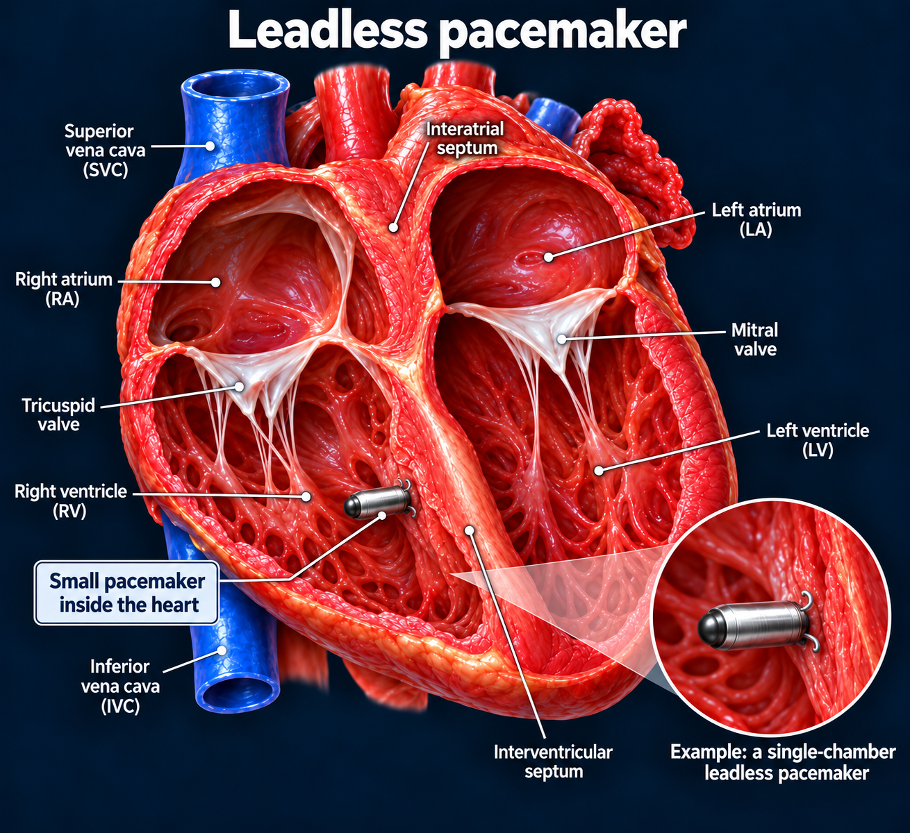
Leadless pacemakers place the pacing device inside the heart without a chest pocket or transvenous lead. Single- and dual-chamber options exist; selection depends on chamber support and other patient factors.[7]
An example single-chamber RV leadless pacemaker. This image does not represent every leadless system.Open full-size image →
Conduction system pacing—His bundle or left bundle branch area pacing—aims to engage native ventricular conduction. It may be chosen to preserve synchrony or reduce pacing-induced cardiomyopathy risk, particularly when frequent ventricular pacing is expected. It is a pacing strategy, not an automatic indication for defibrillation.[6]
Three questions to remember:
Does the patient need reliable pacing? Protection from VT/VF? More coordinated ventricular activation?