Understand pacing indications, how a pacemaker helps, and why the pacing strategy matters.
When does a patient need a pacemaker?
A pacemaker provides electrical support when the heart cannot reliably generate or conduct an adequate rhythm. The indication depends on the mechanism, symptoms, reversible causes, and patient goals—not a heart-rate number alone.
Sinus node dysfunction
Pacing is indicated when symptoms are attributable to sinus node dysfunction after addressing reversible causes. No single heart-rate or pause threshold establishes the indication.
Acquired Mobitz II, high-grade, or complete AV block
Permanent pacing is recommended when the block is not due to a reversible or physiologic cause, even without symptoms.
First-degree or Mobitz I AV block
Pacing is generally considered when symptoms correlate with the block; consider the level of block and progressive conduction disease.
Asymptomatic physiologic or sleep-related bradycardia
Usually does not require pacing. Treat contributing conditions, including sleep apnea when present.
Urgent evaluation comes before a scheduled implant discussion.
New dangerous AV block or unstable bradycardia should follow the emergency/monitored pathway described in Section 2.
How does a pacemaker help?
The device senses intrinsic electrical activity and delivers a small pacing impulse when needed. Programming determines the minimum rate, timing between chambers, and response to activity.[2]
Sinus node dysfunction
Atrial pacing can replace a missing or delayed sinus impulse. With intact AV conduction, that impulse can travel naturally to the ventricles. Rate-responsive programming can help selected patients whose heart rate fails to rise appropriately with activity.
AV block
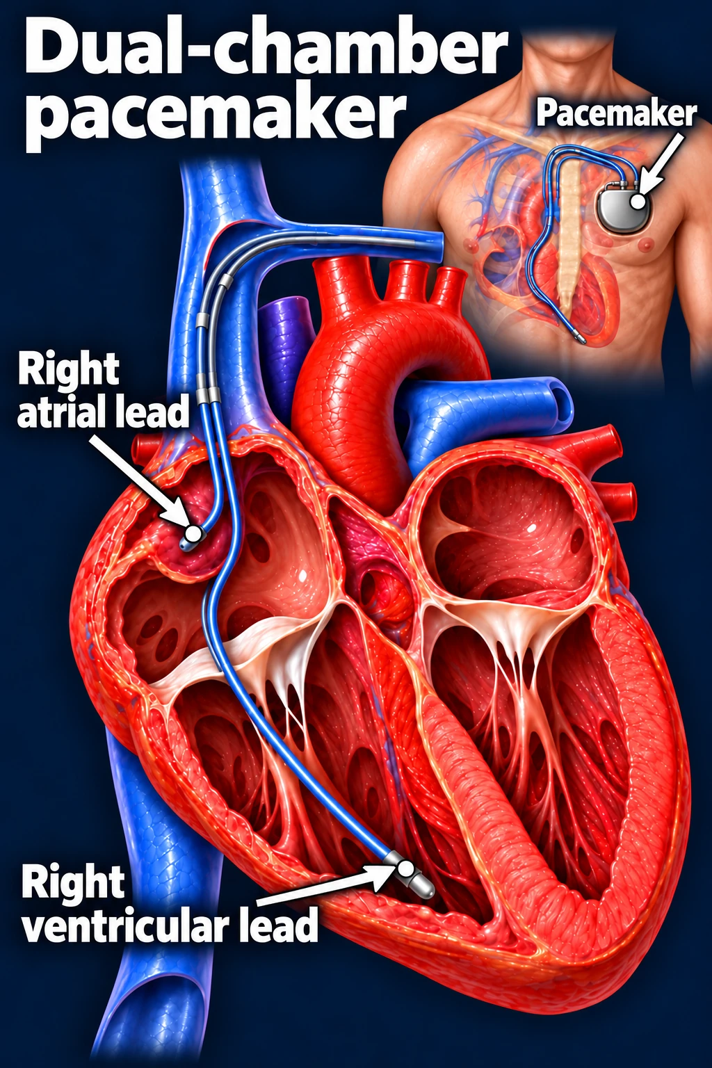
The atrial signal may occur normally but fail to reach the ventricles. A dual-chamber device can sense or pace the atrium and pace the ventricle after an appropriate delay, supporting coordinated activation.
Illustrative conventional dual-chamber pacemaker with an RV apical lead. Lead location and device configuration are individualized; this is not the only pacing approach.Open full-size image →
A pacemaker supports the rhythm; it does not repair the diseased conduction tissue.
It also does not eliminate coexisting AFib or an independent indication for anticoagulation. A standard pacemaker provides pacing rather than defibrillation shocks.
Why pacing location matters
Frequent conventional right ventricular pacing can produce ventricular dyssynchrony and, in some patients, pacing-induced cardiomyopathy. The EP considers ventricular function and anticipated pacing burden when selecting a strategy.
Conduction system pacing—His bundle or left bundle branch area pacing—aims to engage the heart’s conduction system. CRT coordinates ventricular activation through a different pacing configuration. These options help selected patients preserve or improve ventricular synchrony.
For patients with LVEF 36–50% who need substantial ventricular pacing, cardiac physiologic pacing is reasonable to reduce cardiomyopathy risk. It may also be considered in selected patients with normal LVEF and substantial pacing needs. Device selection remains individualized.[3]
What should clinicians explain before referral?
The goal: relieve symptoms caused by bradycardia or protect against unreliable ventricular activation from significant block.
The options: transvenous and leadless systems are available; chamber support, anatomy, and pacing strategy influence selection.
The tradeoffs: discuss infection, bleeding, lead/device problems, and procedure-specific complications with the implanting team.[3]
The ongoing care: scheduled device checks and often remote monitoring assess battery, leads, pacing, and detected rhythms. Batteries eventually require replacement.[4]
Shared decisions should reflect the patient’s preferences, functional goals, overall health, and expected benefit.[1]
After implantation: close the loop
Confirm device-clinic follow-up and remote-monitoring enrollment. Reinforce the implanting team’s wound care, activity, and medication instructions. New syncope, fever, pocket redness/drainage, or worsening symptoms warrants prompt assessment; instability requires emergency care.[4]
Patient explanation: “The pacemaker watches your heart rhythm and supplies an electrical impulse when your own system is too slow or a signal does not reach the lower chambers. The specialist will choose the system that best fits your heart and pacing needs.”