Distinguish impulse generation from conduction failure, compare EKG patterns, and recognize dangerous block.
A slow pulse is a finding—not a diagnosis
Bradycardia describes a slow heart rate. The important question is whether the heart is generating impulses too slowly, failing to conduct them, or responding normally to rest, sleep, or conditioning.
Start with normal conduction
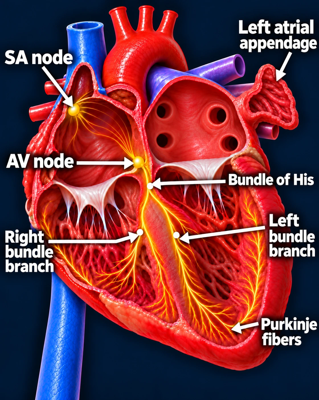
The sinus node starts the atrial impulse. The AV node delays it briefly, then the His–Purkinje system activates the ventricles. On the EKG, the P wave represents atrial activation, the PR interval reflects conduction to the ventricles, and the QRS represents ventricular activation.[2]
Follow one normal heartbeat
SA node → Atria → AV node → Ventricles
The SA node starts the signal. The heart’s natural pacemaker in the right upper chamber begins each normal sinus beat.
The signal spreads through both atria. The upper chambers contract, moving blood into the ventricles.
The AV node briefly delays the signal. This pause gives the ventricles time to finish filling.
The signal activates the ventricles. It travels through the bundle of His, right and left bundle branches, and Purkinje fibers. The lower chambers contract, pumping blood to the lungs and body.
The atria contract first; the ventricles follow. This organized sequence produces a coordinated heartbeat.
The problem is impulse generation or its exit into the atrium. It can cause slow sinus rhythm, pauses, an inadequate rate increase with activity, or tachy-brady episodes. A low heart-rate number alone does not establish the need for a pacemaker; symptom–rhythm correlation is central.[1]
AV block
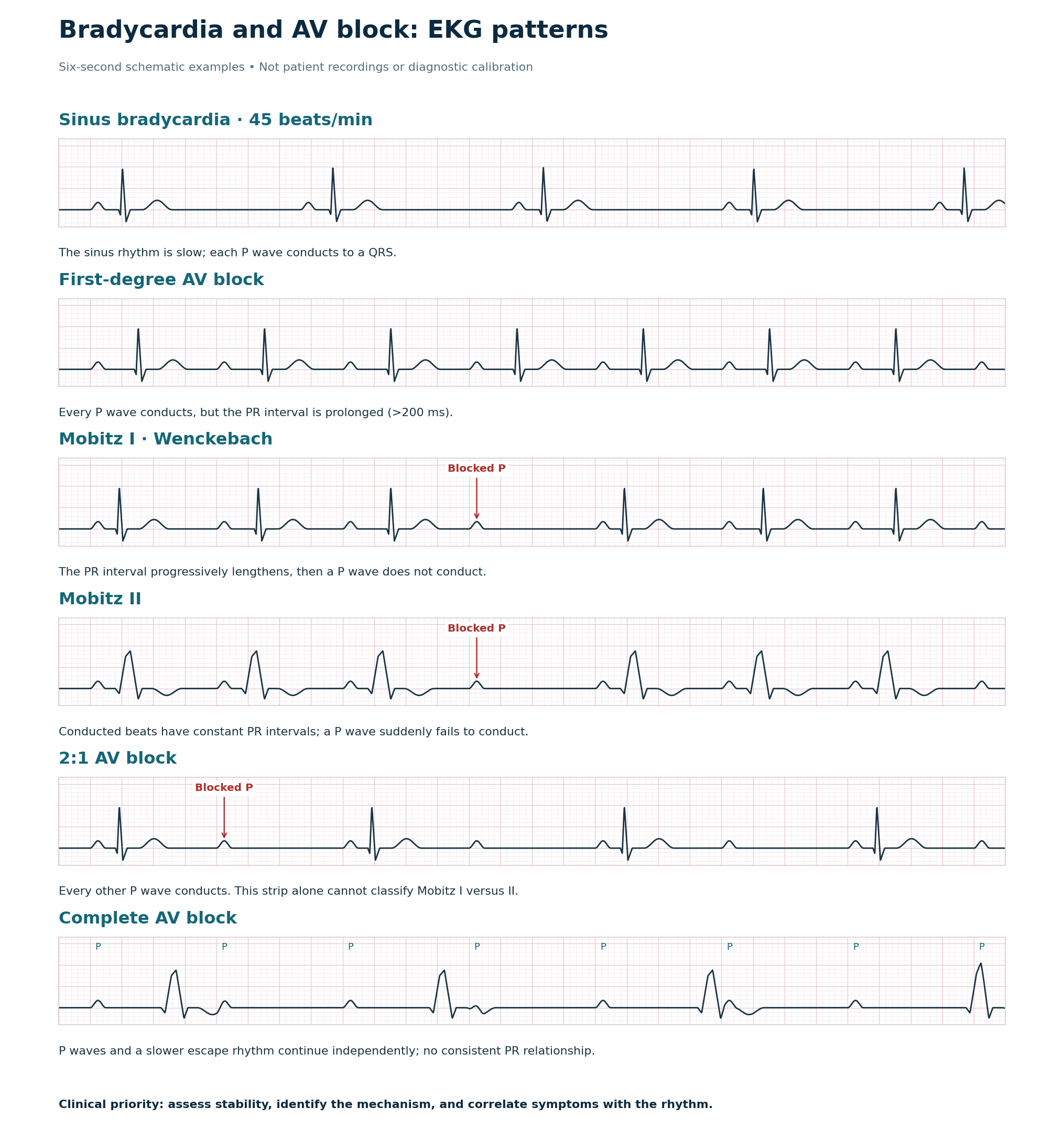
Atrial impulses are delayed or blocked on their way to the ventricles. Look for P waves that continue despite absent QRS complexes. The pattern determines the level of concern.[2]
Compare the EKG patterns
P = atrial activity. QRS = ventricular activity. Schematics for teaching only. In complete block, P waves may overlap QRS/T waves. Wide QRS complexes are shown in the Mobitz II and complete-block examples; QRS width can vary. [1, 2]Open full-size image →
2:1 AV block needs further evaluation: alternating conducted and blocked P waves do not provide enough PR intervals to determine Mobitz I versus Mobitz II.[2]
Why it matters
Bradycardia can cause fatigue, exercise intolerance, dizziness, near-syncope, or syncope. Review rate-slowing medications and reversible contributors before deciding on permanent treatment.[1]
Recognize the urgent patient.
Hypotension, shock, acute altered mental status, ischemic chest discomfort, or acute heart failure with bradycardia requires emergency assessment. In clinic, activate EMS.[3]
New Mobitz II, high-grade, or complete AV block warrants urgent monitored evaluation. Acquired block of these types without a reversible or physiologic cause generally requires permanent pacing regardless of symptoms.[1]
Key distinction: In sinus node dysfunction, symptoms usually drive pacing decisions. Dangerous AV conduction disease may require pacing even when symptoms are minimal.